Download
1 / 51
510 likes | 714 Vues
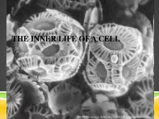
Inner Life of a Cell. Inner Life Of A Cell - Full Version.mkv – YouTube Music Narrative: Harvard http://www.youtube.com/watch?v=GigxU1UXZXo&feature=fvwrel. Agenda: Nov. 26th. Homework: rDNA project due on Friday, Nov 30 th Pfeiffer Thank you notes due on Wed.

E N D
Inner Life of a Cell • Inner Life Of A Cell - Full Version.mkv – YouTube • Music • Narrative: Harvard http://www.youtube.com/watch?v=GigxU1UXZXo&feature=fvwrel
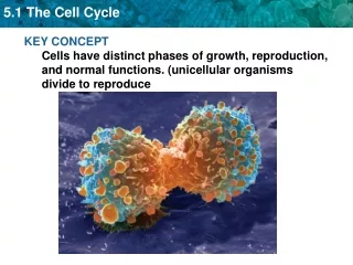
Agenda: Nov. 26th Homework: rDNA project due on Friday, Nov 30th Pfeiffer Thank you notes due on Wed. Objective: To determine how proteins are made Warm up: Central Dogma Proteins and how they are synthesized Gene expression - review of process Shape determines function Senior Project Presentation Tuesday: Proteins in more depth
Warm up: What is the Central Dogma of Biology and Biotechnolgy? Why is it important?
The CENTRAL DOGMAsays: DNA _________ ___________ • The first step will be to convert DNA to ______. This happens in the _______ and the process is called _____________. • Next, the _____ will be converted to a protein. This happens in the ___________ and the process is called ____________. This process will require assistance from the ___________ in the cell.
Transcription and Translation
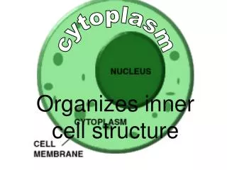
Inside a Cell • Journey Inside The Cell - YouTube
Protein structure and function “Gene Expression” Also known as “how to make a protein and how it works”
What do proteins do? • Each person has 30,000 different types of proteins and many millions of copies. • What is the function of proteins? • Notes for class notebook • Protein Functions in the Body – YouTube • http://www.youtube.com/watch?v=T500B5yTy58
The Structure of Life • Skim pages 6 – 9
Proteins • Structure determines function • Or “proteins are shaped to get the job done” The Structure of Life • Genetic Code p. 12-13 • Peering into Protein Factories p. 23 • Beyond Drug Design pp. 52-55
Proteins • Structure determines function • Or “proteins are shaped to get the job done” • Examples:
Cartilage and tendons 3 strand, rope-like structure provides strength Collagen
Review Base Pairing Rules for Transcription & Translation
See p.12-13 Structure of Life for amino acid names & more details. The Genetic Code
Example of amino acids linked by polypeptide bonds(Note: protein synthesis has direction N to C) Part of a protein: an opioid peptide that modulates the perception of pain
Enzyme: “Pencil transferase” • You will make a new protein (an enzyme) whose job (function) is to transfer a pencil. • Bend the pipe cleaner (chenille stems) so that a pencil can be moved from one table to another. • For the “pencil transferase” to function correctly, you cannot touch the pencil when moving it from your table to the next one. • Keep the successful shape. Draw it in your class notebook.
Questions: Describe the shape of your protein. How would the shape change if one of the amino acids was eliminated?
Proteins – shape determines function Structure: Primary Secondary Tertiary Quaternary
Shape determines function • Primary structure • Order of amino acids • Combine 50-2000 to make proteins • Secondary structure • Alpha helix • Beta Sheet • Plus unstructured loops
Shape determines function • Tertiary • Globular: compact • Fibrous: linear • Quaternary • Multiple polypeptides (amino acid chains) come together
Agenda: Tuesday 11/27 • Read The Structure of Life • The Genetic Code: pp. 12-13 • Worksheet: Genes to Polypeptides • Four Levels of Protein Structure • In more depth • Epigenome – When are proteins produced?
From Genes to Polypeptides • Complete worksheet
See p.12-13 Structure of Life for amino acid names & more details. The Genetic Code
Review: Protein Theater • Setting the scene: • Room walls are the cell membrane • Nucleus • Ribosome • Cytoplasm • Transcription starts with RNA polymerase recognizing a promoter • Gene on the DNA determines the complementary mRNA • mRNA specified the correct sequence for amino acids
Proteins • Structure determines function • Or “proteins are shaped to get the job done” The Structure of Life • Genetic Code p. 12-13 • Peering into Protein Factories p. 23 • Beyond Drug Design pp. 52-55
Differences in the amino acids • Resource: • Chem4Kids.com: Biochemistry:Twenty Amino Acids Amino Acid Sequence of Bovine Insulin
Four Levels of Protein Structure 1. A protein’s primary structure is its amino acid sequence • Primary structure: the sequence of amino acids that form the polypeptide chains • A change in the primary structure can alter the resulting protein
20 amino acids • http://www.personal.kent.edu/~cearley/PChem/amino/3d.htm • http://wbiomed.curtin.edu.au/biochem/tutorials/AAs/AA.html • http://www.chem4kids.com/files/aminoacids/index.html • Chem4Kids.com: Biochemistry:Twenty Amino Acids
Four Levels of Protein Structure 2. Secondary structure is polypeptide coiling or folding produced by hydrogen bonding • Secondary structure: parts of the proteins coil or fold into local patterns • Coiling: alpha helix • Folding: beta pleated sheets
Hydrogen bonds between amino acids • Backbones of the amino acids • C=0 attracted to the NH of the backbone another amino acid • Not the covalent bonds (peptide bonds)
Alpha helix Beta Sheet Secondary Structure – Hydrogen Bonding (See other type.
Secondary shapes often combined into one 3-D structure called a domainEach domain has a function. Note: Also unstructured loops Note: Di-sulfide bridge Strong covalent bond; acts as anchor
3.Tertiary structure is the overall shape of a polypeptide • Tertiary structure: overall 3 dimensional shape of a protein • Globular: compact shape, enzymes • Fibrous: helical, tough, water-insoluble • Result of hydrogen bonding as well as ionic bonding (hydrophilic R groups) • Folded so that hydrophobic R groups are on the inside
Tertiary Structure: hydrophilic vs. hydrophobic R-groups http://www.bio.davidson.edu/courses/genomics/jmol/aatable.html Amino Acid Structures
Basic Rules for Structure based on R groups Hydrophobic Hydrophilic Polar (ionic) Attracted to water since water is polar “Comfortable” in the watery environment of cytosol (cytoplasm) Fold to be on the outside of the protein • Non-polar • R groups with only C& H • Side chains fold up into the interior of the protein
Pipe cleaners Proteins:Shape determined by hydrophilic or hydrophobic • Choose: • 4 pairs of smooth beads • 4 pairs of triangle beads • String the beads in a random order • Triangle beads represent hydrophilic R-groups (same color attracted to each other) • Smooth beads represent hydrophobic R-groups and are in the interior of the protein • Fold the pipe cleaner protein to fit these rules • Draw the shape.
Pipe cleaner proteins • Compare your protein’s shape to others at your table. • How and why are they different? • What conclusions can you make about folding of proteins?
4.Quaternary structure is the relationship among multiple polypeptides of a protein • Quaternary structure: when two or more polypeptide chains come together
Representing the structure of proteins Protein in cell membrane: Left: outside of membrane Purple: where protein crosses Right: inside of cell Receptor protein: pass molecular messages from receptors to inside of cell
Major Unsolved Problem“Protein folding problem” Scientists cannot predict shape & function of a protein based on the gene • Can determine the amino acid sequence • Can now make rough estimates of shapes • Compare to known proteins using data bases (bioinformatics) • Cannot accurately predict the position of each atom