Download

1 / 35
380 likes | 774 Vues
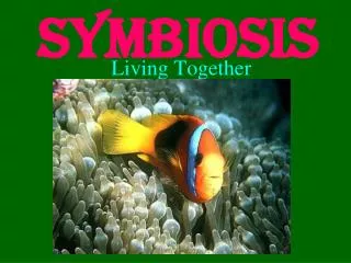
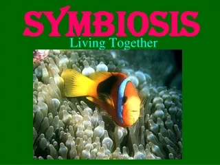
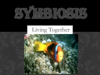
Symbiosis. Sym biosis. together. life. Symbionts : the organisms involved. Host : the larger organism, if there is one. Mutualism : both symbionts benefit. Commensalism : one symbiont receives benefit while neither harming nor helping the other in any significant way.

E N D
Sym biosis together life Symbionts: the organisms involved Host: the larger organism, if there is one Mutualism: both symbionts benefit Commensalism: one symbiont receives benefit while neither harming nor helping the other in any significant way Parasitim: one symbiont, called a parasite, benefits at the expense of the other, usually a host Anthropomorphism in biological definitions?
Examples Ants and plants: mutualism with a large host Epiphytic plants: commensalism with a large host Parasitism: many species of aphids Parasites need to be distinguished between cases where a pathogen may lead to death and where there may be a balance in the host-parasite relationship. For example, there can be genetic balances in virulence and resistance that operate at the populations level. Myxomatosis and rabbits
Symbiosis in the origin of the eukaryotes Two components of the theory 1. The endomembrane may have evolved by infolding of the plasma membrane of an ancestral prokaryote 2. Serial endosymbiosis. Mitochondria and chloroplasts evolved from prokaryotes that lived symbiotically within some ancestral eukaryotes. Molecular evidence – circular DNA in chloroplasts and mitochondria
Nitrogen fixing bacteria In biological nitrogen fixation two moles of ammonia are produced from one mole of nitrogen gas, using 16 moles of ATP and a supply of electrons and protons (hydrogen ions): N2 + 8H+ + 8e- + 16 ATP = 2NH3 + H2 + 16ADP + 16 Pi This reaction is performed exclusively by prokaryotes (the bacteria and related organisms), using an enzyme complex termed nitrogenase. This enzyme consists of two proteins - an iron protein and a molybdenum-iron protein. A point of special interest is that the nitrogenase enzyme complex is highly sensitive to oxygen
Nodule formation 1 Roots emit chemical signals that attract Rhizobium bacteria. The bacteria emit signals that stimulate root hairs to elongate, and to form an infection thread by an invagination of the plasma membrane Soya bean infection with Rhizobium
Nodule formation 2 The bacteria penetrate the root cortex within the infection thread. Plant cells start dividing and vesicles containing the bacteria, bacteriods, bud into the cells from the branching infection thread
Nodule formation 3 Growth continues in the affected regions of the cortex and pericycle and these fuse to form the nodule
Nodule formation 4 The nodule grows and vascular tissue connecting it to the plant’s xylem and phloem develops
Root hairs Lens culinaris (lentil) root nodulation. 7 days after infection 12 days after infection http://www.sunderland.ac.uk/~es0man/tem2.htm
TEM photomicrographs 7 days 12 days Small numbers of bacteria Large numbers of bacteria http://www.sunderland.ac.uk/~es0man/tem2.htm
Clover root with nodules Part of a clover root system bearing naturally occurring nodules of Rhizobium. Each nodule is about 2-3 mm long. http://helios.bto.ed.ac.uk/bto/microbes/nitrogen.htm
Leghaemoglobin In symbiotic nitrogen-fixing organisms such as Rhizobium, root nodules can contain oxygen-scavenging molecules such as leghaemoglobin, which shows as a pink colour when the active nitrogen-fixing nodules of legume roots are cut open. Leghaemoglobin may regulate the supply of oxygen to the nodule tissues in the same way as haemoglobin regulates the supply of oxygen to mammalian tissues Clover root nodules. Leghaemoglobin is found only in the nodules and is not produced by either the bacterium or the plant when grown alone.
Anabaena with heterocysts Anabaena sp. with symbiont bacteria (possibly Zoogloea) around heterocysts In some cyanobacteria nitogen fixation occurs in heterocycts. These cells only have Photosystem I The other cells have both photosystem I and photosystem II, which generates oxygen when light energy is used to split water to supply H2 for synthesis of organic compounds. http://www-cyanosite.bio.purdue.edu/images/images.html
General N fixation and Alfalfa Some legumes are better at fixing nitrogen than others. Common beans are poor fixers (less than 50 lbs per acre) and fix less than their nitrogen needs. Maximum economic yield for beans in New Mexico requires an additional 30-50 lbs of fertilizer nitrogen per acre. However, if beans are not nodulated, yields often remain low, regardless of the amount of nitrogen applied. Other grain legumes such as peanuts, cowpeas, soybeans, and faba beans are good nitrogen fixers, and will fix all of their nitrogen needs. These legumes may fix up to 250 lbs of nitrogen per acre and are not usually fertilized. If large amounts of nitrogen are applied, the plant literally slows or shuts down the nitrogen fixation process. There are many research programs attempting genetic improvement of nitrogen fixation, e.g., alfalfa
Myco rrhizae Fungus Root Mycorrhizas are highly evolved, mutualistic associations between soil fungi and plant roots. The host plant receives mineral nutrients while the fungus obtains photosynthetically derived carbon compounds. Almost 80 percent of all terrestrial plants can form mycorrhizal associations. There are two major types ectotrophic mycorrhizae and endotrophic mycorrhizae
Ectomycorrhizae Mycorrhizal fungi produce a hyphal network in soils consisting of individual strands of hyphae or relatively undifferentiated bundles of hyphae called mycelial strands. Some fungi can produce rhizomorphs, which contain specialised conducting hyphae, or sclerotia, which are resistant storage structures. Soil hyphae acquire nutrients. Fungal structures in soil Absorptive hyphae Mycellial strand Scleridia Mycorrhizal root Rhizomorphs Soil mycellium
Example of ECM short roots (arrows) of birch (Betula alleghaniensis), an angiosperm tree. The mycorrhizal short roots are thicker than other laterals of the same order due to the mantle and Hartig net.
Early stage of colonisation of pine short root by Pisolithus tinctorius. Hyphae (arrows) have contacted the root and are starting to proliferate on its surface near the apex (A). SEM image showing the next stage of pine root colonisation by Pisolithus tinctorius. Mantle hyphae (arrows) have formed a dense covering on the root surface (arrows). http://www.ffp.csiro.au/research/mycorrhiza/ecm.html
Pinus radiata and Amanita muscaria ECM synthesised under sterile conditions. This association has highly branched short roots with many root tips (arrows). Eucalyptus maculata and Astraeus pteridis association synthesised under sterile conditions with relatively unbranched ECM and attached mycelial strands (star).
Hand section cleared and stained with Chlorazol black E and viewed with interference contrast microscopy Populus tremuloides ECM root cross section showing labyrinthine Hartig net hyphae (arrows) around elongated epidermal cells. This complex hyphal branching pattern is considered to increase the fungal surface area in contact with the root. http://www.ffp.csiro.au/research/mycorrhiza/ecm.html
Vesicular arbuscular mycorrhizae The network of hyphae in the soil is only connected to roots by the entry points that initiate mycorrhizal associations Mycorrhizal root system washed carefully from coarse sand to reveal the intact network with external hyphae (arrow) with spores (S) produced by Glomus mosseae. http://www.ffp.csiro.au/research/mycorrhiza/ecm.html
Appressorium At entry point Epidermis Intercellular hyphae in air channel Hypodermis Intracellular hyphae Arbuscules Vesicle Cortex A colony refers to hyphal growth within a root resulting from the same external hyphae (1 or more connected entry points). These are also called infection units.
Arbuscules (A) and convoluted hyphae (arrow) in the inner cortex of an Asarum canadense root. Arbuscules only form in the innermost cortex cell layer next to the endodermis in this species. Vesicles (V) produced by a Glomus species in a leek root. This root also contains many intercellular hyphae. (Bar = 100 um) http://www.ffp.csiro.au/research/mycorrhiza/ecm.html
Lichens Lichens are symbiotic associations between photosynthetic micro-organisms held in a mesh of fungi. Cladonia fimbriata Different species of lichens growing on a boulder. The fungus gives the lichen its shape. The alga provides the fungus with food. In some lichens the ‘alga’ is a Cyanobacteria and fixes nitrogen. Lichens are colonizers of bare rock but some species are epiphytes found in tree canopies.
There are some 16,000 species of lichen The algal components can live in isolation with the fungal component – so the symbiotic relationship is not obligate for them.However, no zoospores (the algal resting stage) or gametes are produced while the alga is in the symbiotic relationship though they can produce them when cultured outside of it. In contrast the symbiotic relationship is obligate for the fungal component. The mycelium of a few species may grow under laboratory conditions, on agar, but no lichen specific thallus is produced and no fruiting body develops. In contrast to the many thousands of lichen fungi, there are only about 100 photosynthetic partners. The oldest certain fossil lichen is EarlyDevonian (about 400 million years old) from the Rhynie Chert.
Cross-section through a lichen Fruiting body of the fungus Fungal hyphae: a filament, composed of single cells joined together end to end See also Fig. 17.18B (but note names used here)
Xanthoria parietina The foliose lichen Xanthoria parietina, which grows on surfaces, including concrete, and rocks subjected to sea spray. Much of the surface is covered with bright orange fungal fruiting bodies about 3-5 mm diameter (arrowheads). The orange colour is due to production of the pigment parietin at the lichen surface. Cross section of one of a lobe viewed by phase-contrast microscopy. The photosynthetic zone (p) is a distinct band of green algal cells. Above this band is the cortex (c) of densely packed fungal cells . The lower part of the thallus consists of a medulla (m) with conspicuous air pockets (a), and a thin lower cortex (lc). Like many foliose lichens, Xanthoria produces rhizinae (r) that penetrate into crevices and help to anchor the lichen to a surface. http://helios.bto.ed.ac.uk/bto/microbes/lichen.htm
Classification by form Lichens are unique as a symbiotic relationship because they look and behave quite differently from their component organisms. Lichens are regarded as organisms in their own right and are given generic and species names. The fungus is termed the mycobiont The green alga or a cyanobacterium is termed the photobiont. The "body" of a lichen is termed the thallus, and its general shape is used to group lichens into four broad categories. FolioseFruticoseSquamuloseCrustose
Foliose lichens Foliose lichens have a flat, leaf-like structure Parmelia physodes, growing on the twigs of a shrub.
Fructicose and Squamulose Fruticose lichens have an erect or pendulous, bushy structure Squamulose lichens have a thallus consisting of minute, scale-like squamules Lecanora muralis with ascocarps pink-brown
Crustose lichens Crustose lichens produce a flat crust on or beneath rock or tree surfaces
Trebouxia Trebouxia The most common photobiont is a single-celled green algae of the genus Trebouxia found in many lichens of temperate, arctic and alpine regions, including all species of the common lichen genus Cladonia. Trebouxia species seldom grow as free-living cells in nature but seem to be specialised lichen symbionts. Trebouxia secretes about 8% of the carbohydrates it produces when living free in experiments, but up to 40% when it is a photobiont. The increased secretion rate is caused by the fungus. In lichens with Trebouxia the fungal hyphae can produce short branches that penetrate through the algal wall to serve as nutrient-absorbing haustoria.
Lichens are remarkable for their ability to withstand prolonged drying and to resume activity rapidly after rewetting. The rewetting of lichens is thought to start by water absorption in the gelatinous matrix of the cortex. The maximum rates of nutrient release from the photobiont occur in optimal moisture conditions, whereas it retains most of the carbohydrate in conditions of water stress. It has been suggested that cycles of wetting and drying may be advantageous in maintaining a lichen symbiosis, because both partners could gain sufficient carbohydrate at different stages of this cycle. They are extremely intolerant to SO2
Sections you need to have read 16.20 17.18 32.11 36.5 Courses that deal with this topic Botany 371/372 Plant physiology laboratory