
Low-Frequency rTMS and Brain Metabolism in Auditory Hallucinations: A PET Imaging Study
This study investigates the impact of low-frequency repetitive transcranial magnetic stimulation (rTMS) on regional brain metabolism, particularly in patients experiencing medication-resistant auditory hallucinations. Utilizing 18FDG PET imaging, we confirm increased metabolic activity in the left temporo-parietal cortex associated with auditory hallucinations. Our results indicate that rTMS effectively reduces metabolism in this region, alongside improved clinical symptoms. We propose an algorithm for individualized neuronavigated rTMS treatment based on specific PET analysis for targeted therapy.

Low-Frequency rTMS and Brain Metabolism in Auditory Hallucinations: A PET Imaging Study
E N D
Presentation Transcript
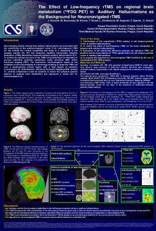
B A MRI Functional neuroimaging in neuronavigated TMS The Effect of Low-frequency rTMS on regional brain metabolism (18FDG PET) in Auditory Hallucinations as the Background for Neuronavigated rTMS J. Horacek, M. Brunovsky, M. Klirova, T. Novak, L. Skrdlantova, M. Kopecek, F. Spaniel , C. Hoschl Prague Psychiatric Centre, Prague, Czech Republic Centre Of Neuropsychiatric Studies, Prague, Czech Republic Third Medical Faculty Of Charles University, Prague, Czech Republic Aims of the study 1) Confirmation of the overactivity (18FDG uptake) in left temporo-parietal cortex in auditory hallucinations. 2) To detect the effect of low-frequency rTMS on the brain metabolism in patients with auditory hallucinations. 3) Evaluation of the anatomical difference between the standard rTMS coil position and the local maxima of 18FDG uptake within the left temporo-parietal cortex in auditory hallucinations. 3) To propose the algorithm for neuronavigated rTMS treatment by the use of individualized PET SPM analysis. Materials and Methods Samples: 12 patients (5F/ 7M) with paranoid schizophrenia (DSM IV, mean age= 34.4, SD=9.1, mean duration of SCH=76.3 months, SD=47.0) with medication-resistant auditory hallucinations (10). Stabile antipsychotic medication for at least 3 weeks. 20 controls (10F/10M, mean age=34.4,SD=9.1). The regional brain metabolism:18FDG PET in Random Episodic Silent Thinking codition, ECAT EXACT 922 (CTI/Siemens, Knoxville, TN) PET scanner. Scans were normalized and smoothed with (12 mm). Statistical Parametric Mapping (SPM99). Low-frequency rTMS (0.9Hz, 100% of motor threshold, 20 min.) applied to the left temporo-parietal cortex for ten days in the treatment of medication-resistant auditory hallucinations. Introduction Neuroimaging studies indicate that auditory hallucinations are associated with the overactivity of the auditory-linguistic cortex [1-4]. Low-frequency rTMS (≤1 Hz) inhibits the cortical excitability and leads to a weakening of the synaptic transfer [5]. The low frequency repetitive transcranial magnetic stimulation (rTMS) diminishes treatment-resistant auditory hallucinations (6,7). To achieve the optimal accuracy of the rTMS coil positioning it is necessary to process individual anatomic parameters using structural (MR) and/or functional imaging (PET). The stereotactic neuronavigation based on the evaluation of the neuroimaging is a technology enabling to target the rTMS coil over a dysfunctional cortical region with a high degree of anatomic accuracy(mm) (8). Individualized Statistical Parametric Mapping-SPM (patient vs. controls comparison) could be sensitive to detect the symptom specific patterns of regional brain metabolism and represents a useful tool for neuronavigation (9). Results: Figure 2: The influence of low frequency 0.9 Hz rTMS on regional brain metabolism. The most pronounced decreased of 18FDG PET uptake after rTMS treatment was found in the left temporal cortex and in the cerebellum (A). 18FDG PET uptake following low frequency rTMS increased (B) in the middle frontal gyrus bilaterally and in the right temporo-parietal cortex. Note: R, right hemisphere; L, left hemisphere, T=4.14, p 0.001. After 2 weeks of the treatment, we found a significant decrease in the total and positive symptoms (PANSS), and on the hallucination scales (HCS, AHRS) (p0.05, data not shown). Figure 1: The higher regional brain metabolism in patients with schizophrenia (N=12) in comparison with controls (N=20). The metabolism is increased in the L superior, middle and inferior temporal gyrus, R and L putamen, R middle occipital gyrus, claustrum and cuneus and cingulate (T = 5.35, p0.05 corr.). Arrow indicates the standard position of rTMS coil (midway between the T3 and P3 sites according to the international 10/20 EEG electrode system). L R L R Figure 4: The proposed algorithm for the neuronavigated rTMS treatment based on the individualized SPM analysis of 18FDG uptake Figure 3: The difference between standard position of rTMS coil (B) over the left T-P cortex (the midway between the T3 and P3 sites according to the international 10/20 EEG system) and local maximum of 18FDG uptake in individualized SPM (A). The MRI and SPM contrasts were coregistered in 3D native space (p0,05). For the subgroup of 6 patients the difference is 28.2 mm (s.d.=7.0) 1a 18FDG PET Preprocessing: . (Normalization and smoothing) Patient with auditory hallucinations 1b 5a 5b Different modalities of data integrated in neuronavigation: a) MRI b) SPM PET contrast c) individual brain anatomy d) head position e) known PET correlates of symptoms (a priori) 2 Individualized SPM: Comparison with controls One sample t-test p0,05 (9) 4 6 3 SPM contrast (1 -1) Coregistration with native (non-normalized) MRI • Discussion: • Our findings confirm the increased metabolism in the left temporo-parietal cortex in auditory hallucinations. • The rTMS decreased the brain metabolism in the left superior temporal gyrus and in interconnected regions and effected increases in the contralateral cortex and PFC • We detected the substantial difference between standard rTMS coil position and the local maximum of metabolism in individualized SPM. • The rTMS neuronavigated according to the local maxima of 18FDG uptake is very promising possibility for more pronounced clinical effect. • The algorithm for neuronavigated rTMS is proposed and would be tested in a controlled trial. Acknowledgment: The research was supported by the projects he projects 1M0517 MSMT CR and NR8792 of IGA MZ CR. References: (1)Copolov DL, et al. Cortical activation associated with the experience of auditory hallucinations and perception of human speech in schizophrenia: a PET correlation study. Psychiatry Res 2003;122:139-152. (2) Suzuki M, et al. Left superior temporal blood flow increases in schizophrenic and schizophreniform patients with auditory hallucination: a longitudinal case study using 123I-IMP SPECT. Eur Arch Psychiatry Clin Neurosci 1993;242:257-261. (3) Shergill SS, et al. Mapping auditory hallucinations in schizophrenia using functional magnetic resonance imaging. Arch Gen Psychiatry 2000;57:1033-1038. (4) Volkow ND, et al. Phenomenological correlates of metabolic activity in 18 patients with chronic schizophrenia. Am J Psychiatry 1987;144:151-158. (5) George MS et al. Mechanisms and state of the art of transcranial magnetic stimulation. J ECT 2002;18:170-181. (6) Hoffman RE et al. Transcranial magnetic stimulation of left temporoparietal cortex and medication-resistant auditory hallucinations. Arch Gen Psychiatry 2003;60:49-56. (7) Lee SH et al. A double blind study showing that two weeks of daily repetitive TMS over the left or right temporoparietal cortex reduces symptoms in patients with schizophrenia who are having treatment-refractory auditory hallucinations. Neurosci Lett 2005;376:177-181. (8) Hoffman RE et al. Temporoparietal transcranial magnetic stimulation for auditory hallucinations: safety, efficacy and moderators in a fifty patient sample. Biol Psychiatry 2005;58:97-104. (9) Herwig U, Kolbel K, Wunderlich AP, Thielscher A, von Tiesenhausen C, Spitzer M, Schonfeldt-Lecuona C: Spatial congruence of neuronavigated transcranial magnetic stimulation and functional neuroimaging. Clin Neurophysiol 2002;113:462-468.(10) Kopeček M et al. Regional cerebral metabolic abnormalities in individual patients with nonquantitative 18FDG PET and qEEG (LORETA). Psychiatrie 2005;9:56-63.


