Acknowledgements
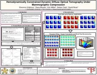
Parameter. Value. Background OC. 0.448 µmol/L/s. Tumor OC. 0.672 µmol/L/s. Background blood flow,. 0.000275 L/L/s. Tumor blood flow,. 0.0006875 L/L/s. Total tissue hemoglobin 10% change over 60sec. a = 18 µmol/L b = a/600. Total blood hemoglobin, HbTbl. 700 µmol.

Acknowledgements
E N D
Presentation Transcript
Parameter Value Background OC 0.448 µmol/L/s Tumor OC 0.672 µmol/L/s Background blood flow, 0.000275 L/L/s Tumor blood flow, 0.0006875 L/L/s Total tissue hemoglobin 10% change over 60sec. a = 18 µmol/L b = a/600 Total blood hemoglobin, HbTbl 700 µmol Arterial blood oxygen saturation, SaO2 0.98 Initial background oxygen saturation, SO2.init 0.7 (70%) Initial tumor oxygen saturation, SO2,init 0.85 (85%) |SO2original – SO2ranging| 0 1 0.0003 0.5 OC 0.00015 F OC and Flow reconstruction when 60dB SNR used in data generation OC and Flow reconstruction when no noise used in data generation OC reconstruction if we use the tumor flow value (60dB SNR for generated data) OC reconstruction if we use the background flow value (60dB SNR for generated data) Hemoglobin Time Curves Hemodynamically Constrained Dynamic Diffuse Optical Tomography Under Mammographic CompressionEleonora Vidolova1, Dana Brooks1, Eric Miller2, Stefan Carp3, David Boas31. Northeastern University; 2. Tufts University; 3. Massachusetts General Hospital; 1. Introduction 3. Reconstruction • DOT taken along x-ray mammogram provide valuable functional information. • X-ray mammography only gives structural information → hard to distinguish between benign and malignant masses. • Oxygen consumption (OC) and blood flow (F) contrasts observed between malignant and normal tissue[3, 4]. • Blood flow, oxygen saturation(SO2), water and lipid distributions could specify the degree of malignancy of a tumor. • Physiological changes in the breast due to mammographic compression are significant → should be taken into account[2]. Invert data using Tikhonov regularization • Hemodynamic model describing the relation between hemoglobin content, OC, SO2 and F in the breast during mammographic compression. • OC and F could become novel breast cancer optical markers [2]. • Indirect method: First reconstruct hemoglobin content and then OC and F. 2. Forward Model and Simulation Data • Generated data for background and tumor regions using equation (2), representing 90s of mammographic compression at 6 lbs • Mass balance of HbO within a volume gives [2], • Where, SaO2 and SvO2 are arterial and venous oxygen saturation; OC is oxygen consumption, F is blood flow and V is volume. • Factor of 4 accounts for 4 O2 molecules bound to each Hb molecule. (1) Parameter Fitting - Oxygen Consumption and Blood Flow Reconstruction • Assumptions: • SO2 = mean (SaO2, SvO2) • SO2 =HbO/HbT, with initial condition, SO2,init • HbTtissue = a+bt (Clinical data suggest that HbT changes over time, previous research assumed HbT constant [1,2]) (2) • Fitting of OC and Flow is very dependent on the SNR of the generated data • If fitting for OC only we get a better fit if we know which region we are in 4. Future Work • Compare reconstruction results to the constant HbT model used before • Analyze how small perturbations in the reconstructed HbO and HbR affect the oxygen consumption and blood flow reconstructions • Use the reconstructed SO2 data do differentiate between regions and use that information when fitting for oxygen consumption Forward Data • Added electronic noise, modeled as i.i.d Gaussian random variables. • Simulations done in PMI toolbox developed at MGH. Acknowledgements References • This work was supported by CenSSIS, under the Engineering Research Centers Program of the National Science Foundation (Award # EEC-9986821). • D. Boas and S. Carp acknowledge support from NIH grants R01-CA97305 and 54-CA105480. • This work is a continuation of the work done by Dibo Ntuba on her Masters degree project. • D. T. Ntuba, S. A. Carp, G. Boverman, E. L. Miller, D. A. Boas, D. H. Brooks. “Reconstructing oxygen consumption and blood flow in diffuse optical tomographic breast imaging under mammographic compression.” CenSSIS RICC 2006 Student Poster Session. • S. A. Carp, T. Kauffman, Q. Fang, E. Rafferty, R. Moore, D. Kopans, D. Boas. “Compression-induced changes in the physiological state of the breast as observed through frequency domain photon migration measurements.” Journal of Biomedical Optics, Vol. 11(6), Nov./Dec. 2006 • 3. T. Durduran, R. Choe, G. Yu, C. Zhou, “Diffuse optical measurement of blood flow in breast tumors”, Optics Letters 30(21), 2915-17 (2005). • R. Beaney, A. Lammertsma, T. Jones, C. Mckenzie, K. Halnan, “Positron emission tomography for in-vivo measurement of regional blood flow, oxygen utilisation, and blood volume in patients with breast carcinoma”, The Lancet, 1(8369), 131-134 (1984).