Download
1 / 70
751 likes | 1.45k Vues
Principles of Radiographic Interpretation Juan F. Yepes DDS , MD, MPH Assistant Professor Division of Oral Diagnosis, Oral Medicine and Oral Radiology University of Kentucky, College of Dentistry Spring 2009. What is that radiologist saying?. Well defined and Corticated….. .

E N D
Principles of Radiographic Interpretation Juan F. Yepes DDS, MD, MPH Assistant Professor Division of Oral Diagnosis, Oral Medicine and Oral Radiology University of Kentucky, College of Dentistry Spring 2009
What is that radiologist saying? Well defined and Corticated…..
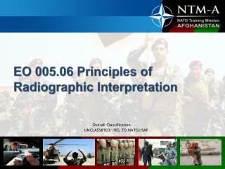
General Objective • The objective of this lecture is to provide step-by-step- analytic process • that can be applied to the interpretation of diagnostic images. • Proficiency comes with PRACTICE!!! • Radiographic interpretation of caries • Radiographic interpretation of periodontal disease • Radiographic interpretation of benign conditions • Radiographic interpretation of malignant conditions
Radiopaque This refers to the item that is being imaged, i.e. in our case a part of the patient, and means that it blocks the transmission of x rays.
Radiolucent This refers to the item that is being imaged, i.e. in our case a part of the patient, and means that it permits the transmission of x rays.
Dense (Density) In radiology this usually refers to the film, and refers to the ability of the film to block the transmission of light (i.e. blackness)
Well localized The item being reported is limited to a specific area, and does not extend beyond that locality.
Poorly localized The item being reported is not limited to a specific area, and extends into surrounding anatomical sites.
Well defined The edges of the item being reported are reasonably sharp and clearly define the extent of the lesion.
Poorly defined The edges of the item being reported are not sharp. The actual borders and thus the exact extent of the lesion are not clearly defined.
The lesion may thus be well localized and well defined or well localized, but poorly defined or poorly localized and poorly defined but generally not poorly localized and well defined
Corticated The entity being reported is not only well defined, but has a cortex, i.e. an osseous border, seen as a thin white line.
Multilocular The entity being reported is usually well defined and has a cortex, i.e. an osseous border, seen as a thin white line, but is partially or totally subdivided into several loculi.
Multilocular Loculus, loculi: the diminutive of locus. Locus, loci: a place or position
Multilocular Thus, multilocular implies several small places. As we use it, they are joined places.
Osteitis vs Osteomyelitis Both terms mean that there is inflammation of bone.
Osteitis inflammation of bone, involving the haversian spaces, canals, and their branches, and generally the medullary cavity, and marked by enlargement of the bone, tenderness, and a dull aching pain. Dorland’s Illustrated Medical Dictionary 29th ed.
Osteomyelitis inflammation of bone caused by infection, usually by a pyogenic organism, although any infectious agent may be involved. It may remain localized or may spread through the bone to involve the marrow, cortex, cancellous tissue, and periosteum. Dorland’s Illustrated Medical Dictionary 29th ed.
Osteitis inflammation of bone that remains localized, and may be more of a painful inconvenience
Osteitis Rarefying Osteitis Sclerosing Osteitis
Rarefying Osteitis(Periapical lesion U. of K.) Inflammation of bone that results in the removal of bone. The term is not a diagnosis, but a radiologic interpretation that includes abscess, cyst and granuloma.
Sclerosing Osteitis I use the term sclerosingosteitis, i.e. inflammation of bone (osteitis) that causes sclerosis.
Periosteal Reaction Any involvement of the periosteum by a pathological process that results in the deposition of periosteal new bone.
Inflammation of the Jaws and Periosteal Reactions Sessile Osteomyelitis Periostitis Healing Orthogonal Anemia Sarcoma Sarcoma
Periapical Radiolucency This is merely a description of a finding, and should better be stated as a periapical radiolucent area or line. It does not denote disease. e.g. the maxillary sinus could be a periapical radiolucent area, as could the mental foramen.
What is that radiologist really saying? Well defined and Corticated
Well localized The radiologist infers that the appearance is consistent with a slow non-invasive growth, and thus that this is benign.
Poorly localized The radiologist infers that the appearance is consistent with a faster and invasive growth, and thus that this is malignant, or a spreading infectious/inflammatory lesion.
Well defined The radiologist infers that the appearance is consistent with a slow non-invasive growth, and thus that this is benign.
Poorly defined The radiologist infers that the appearance is consistent with invasive growth, and thus that this is malignant, or infectious/ inflammatory lesion.
Corticated The radiologist infers that the appearance is consistent with a slow non-invasive growth, and thus that this is benign.