Addressing EU Social Challenges in Medical Biotechnology Education at Pécs and Debrecen Universities
170 likes | 330 Vues
This document explores the incorporation of contemporary social challenges faced by the European Union into the medical biotechnology master's programs at the University of Pécs and the University of Debrecen. It highlights the importance of adapting educational materials to meet evolving societal needs, particularly in areas like transdifferentiation and regenerative liver medicine. The paper, authored by Dr. Péter Balogh and Dr. Péter Engelmann, delves into liver regeneration processes, stem cell applications, and the clinical necessities surrounding organ transplantation.

Addressing EU Social Challenges in Medical Biotechnology Education at Pécs and Debrecen Universities
E N D
Presentation Transcript
Manifestation of Novel Social Challenges of the European Unionin the Teaching Material ofMedical Biotechnology Master’s Programmesat theUniversity of Pécs and at the University of Debrecen Identificationnumber: TÁMOP-4.1.2-08/1/A-2009-0011
Manifestation of Novel Social Challenges of the European Unionin the Teaching Material ofMedical Biotechnology Master’s Programmesat theUniversity of Pécs and at the University of Debrecen Identification number: TÁMOP-4.1.2-08/1/A-2009-0011 Dr. PéterBalogh and Dr. Péter Engelmann Transdifferentiation and regenerative medicine – Lecture 8 Liverregenerationfromstemcells
Structure of thehepaticlobe Bilecanaliculi Centralvein Centralvein Portaltracts (triads) Sinusoids Bileduct Portaltract Branchportal vein Branchhepatic artery Periportal (6-8 cells) Centrilobular (8-10 cells) Glutaminesynthetase + (1-3 cells) Limiting plate
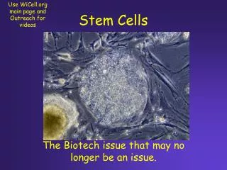
Clinical necessity of liver regeneration • Shortage of livers for orthotopic liver transplantation • Liver cell transplantation – limited amount • Choice of stem cell candidates – variable success in experimental conditions
Main phases of liverregeneration 1 Migration 3 Clearance Physical/chemical/geneticalstimulus Gadoliniumchloride/ monocrotaline Organdamage Deadcell MMP-9 Centralvein Kupffercells (phagocytosis) Centralvein SDF-1 HGF (SCF) Recruitment Stemcells (c-kit, c-met, CXCR4) Immunosuppression Encapsulation Co-transplantation Effectorcells Monocrotaline Doxorubicin Hepaticinjury VEGF 2 Integration VEGF HGF TGF FGF Centralvein Organdamage Alteration of blood flow Vasodilatators Sinus endothel permeability Gapjunctions Variablein vivo cellphenotype MMP-9 MMP-2 MT1-MMP Cellloss of 70-80%
Developmentalrelationshipbetweenhepatic-pancreaticdifferentiationDevelopmentalrelationshipbetweenhepatic-pancreaticdifferentiation ? Ovalcellprogenitor Pancreatic progenitor(s) Hepatic oval cell Pancreatic oval cell Bile duct Hepatocyte Endocrine cell Pancreatic duct Acinar cell
Transcriptional control of hepatoblast development Hepatoblast HGF C/EBP HNF-6 Tbx3 HNF-1 Wnt BMP+FGF FoxM1B ECM HNF-4 Notch2 Cholangiocyte Hepatocyte Albumin ? ECM C/EBP Hex Jagged Core transcription factor network: HNF-6/OC-2 TGF HNF-4 HNF-1 HNF-6 Parenchyma Periportal HNF-1 LRH-1 Foxa2 Sox9 HNF-1 Hepatocyte maturation cords Cholangiocyte maturation ducts
Oval cells – adult liver stem/progenitor cells • Origin: debated (their precursors are associated with the biliary tree) • Bipotential differentiation: hepatocyte and cholangiocyte • Phenotype: shared markers with adult hepatocytes (albumin, cytokeratins 8 and 18), bile duct cells (cytokeratins 7 and 19, OV-6, A6), fetal hepatoblasts (AFP), and haematopoietic stem cells (Thy -1, Sca-1, c-kit).
Cellular targets for hepatic regeneration • Hepatocytes: metabolic activity of the liver • Cholangiocytes: formation of bile ducts • Both derive from embryonic endodermal epithelium.
Stages and forms of liver regeneration • Surgical partial hepatectomy – from hepatocytes (often polyploid cells) • Possible sources: hepatocytes, oval cells and extrahepatic stem cells (HSC?) • Assessment of lineage commitment: albumin, glucose-6-phosphatase, transferrin and transthyretin (hepatic). • Fibrotic regeneration: transformation of fibrocytes into myofibroblasts • Parenchymal regeneration: regeneration of hepatocytes
Sequence of parenchymal regeneration of the liver • Stem cell migration into the liver parenchyma is directed by chemoattractive agents (as SDF-1, HGF and SCF) secreted by damaged liver cells • Increased MMP-9 expression by host hepatocytes after injury, leading to ECM remodeling and increased vascular permeability • Transformation of local microenvironment for the integration and proliferation of the transplanted cells, including local secretion of cytokines/growth factors (HGF, FGF, TGFa). Dead cells will be phagocyted by Kupffer cells.
Oval cell activation and expansion • Liver injury activates oval cells (their precursors in the biliary tree?) AND other support cells (stellate cells, macrophages/Kupffer’s cells, NK cells, endothelium, etc) • Homing/intrahepatic migration to the site of injury • Proliferation and bidirectional differentiation (hepatocyte/cholangiocyte)
Non-hepatic cells for liver regeneration • Autologous: Bone marrow-derived/mesenchymal stem cells – fibroblastic regeneration • Allogenic: Fetal-derived hepatocytes or embryonic stem cell-derived liver cells
Differentiation of iPS cells into hepatocytes • Induction of iPS cells: transfection with TFs • Formation of embryoid bodies • Induction of endodermal commitment: treatment with Activin A and bFGF • Differentiation into hepatocytes: treatment with hepatocyte growth factor (HGF) • Assessment: gene expression, albumin secretion, glycogen storage, urea production, and inducible cytochrome activity
Summary • Depending on the origin/type of liver damage, different regeneration processes operate, thus (a) in loss of liver mass, the regeneration is initiated from hepatocytes, whereas (b) in toxicity from hepato-cholangiocyte progenitors. • Oval cells as adult-type hepatocyte/cholangiocyte progenitors are most likely to be facultative stem cells, although cells with stem cell activity from extrahepatic sources may also operate in liver regeneration.