Download
1 / 9
100 likes | 335 Vues
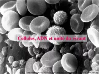
Cellules, ADN et unité du vivant. http://www.molecularstation.com/molecular-biology-images/data/504/482px-SEM_blood_cells.jpg. Méthode : Comment utiliser un microscope optique. 1. Légender le dessin du microscope optique . 2. Placer dans l’ordre et en les complétant les phrases suivantes.

E N D
Cellules, ADN et unité du vivant http://www.molecularstation.com/molecular-biology-images/data/504/482px-SEM_blood_cells.jpg
Méthode : Comment utiliser un microscope optique 1. Légender le dessin du microscope optique 2. Placer dans l’ordre et en les complétant les phrases suivantes (vous pouvez utiliser plusieurs fois la même phrase) 7 oculaire 2 3 objectif 4 platine 1 diaphragme 9 lumière vis micrométrique 6 6/8/10 6/8 vis macrométrique 5 réglage de la lumière Ou intérupteur 11 huile
x 4 x 40 x 10 4 mm 1,5 mm x 100 x 10 x 10 x 400 x 40 x 10 0,5 mm x 10 x 1000 x 100 Grossissement = objectif x oculaire
Microscope électronique http://www.royalbcmuseum.bc.ca/exhibits/journeys/francais/print/forest_3_1c.html http://www.molecularstation.com/fr/molecular-biology-images/showphoto.php/photo/25/limit/recent
Épiderme buccal, d’un être vivant pluricellulaire Epiderme de grenouille http://microscopique.blogspot.com/2007/06/epiderme-de-grenouille.html http://homepage.mac.com/danielbalas/HISTOLOGIE/EPITHDIG/cbgsoe/cbgsoe14.jpg
Un être vivant unicellulaire : la paramécie http://membres.lycos.fr/nabiga/animaux/ciliophores.html
Champignon de Paris Cèpes http://www.kamaxx.com/jdlf/img/photos/4210_1.jpg http://www.gitescantal.fr/images/photos/cepe.jpg Morilles http://richardg.blogs.com/photos/uncategorized/2007/04/22/morille_grise_2.jpg
Cellules végétales non chlorophylliennes Plante non chlorophyllienne : Cellules d’oignon L’Orobanche, une plante parasite Cellules de tomate Cellules de racine doc18 p163 http://alain.granier2.free.fr/vsp/microbes/cellules_vegetales_et_animales.htm http://www.regione.calabria.it/ambiente_1/Julia%20Sila/Progetto%20Sila%20Greca/laflora5.htm
A l’aide des photos du livre, remplir le tableau suivant : Champignon unicellulaire Bactérie unicellulaire Végétal pluricellulaire chlorophyllien Végétal pluricellulaire non chlorophyllien 30 µm 60 µm 75 µm 3 µm 2 µm Noyau Mitochondrie Noyau Mitochondrie Vacuole Chloroplaste Noyau Mitochondrie Vacuole Noyau Mitochondrie Eucaryote Eucaryote Eucaryote Procaryote Eucaryote