Heart failure
720 likes | 751 Vues
Heart failure, also known as congestive heart failure, is a common condition that affects millions globally. Explore the causes, types, and clinical manifestations of left-sided and right-sided heart failure along with insights on ischemic heart disease and its clinical syndromes.

Heart failure
E N D
Presentation Transcript
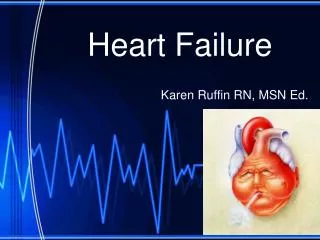
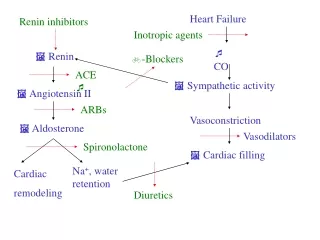
Heart failure • Heart failure (also called congestive heart failure, or CHF) is a frequent end point of many of the conditions • In the United States alone, CHF affects nearly 5 million individuals annually, necessitating >1 million hospitalizations, and contributes to death of 300,000 patients a year.
Most heart failure is the consequence of systolic dysfunction, the progressive deterioration of myocardial contractile function.
Causes of CHF • 1-ischemic heart disease • 2-hypertension. -in 20% to 50% of patients the heart contracts normally but relaxation is abnormal ("diastolic" failure ). -the patients with "diastolic" failure are generally older and more likely to be female with hypertension or diabetes mellitus.
3-valve failure (e.g., endocarditis) • 4-normal hearts suddenly burdened with an abnormal load (e.g., fluid or pressure overload). • 5-acute blood loss
As a compansation the heart dilates the ventricular wall tension increases which increases the oxygen requirements of an already compromised myocardium.
With time, the failing myocardium is no longer able to propel sufficient blood to meet the needs of the body, even at rest. • At this point, patients enter a phase termed decompensated heart failure
Types of Heart failure: • 1- predominantly the left side • 2- predominantly the right side • 3- both sides of the heart.
The most common causes of left-sided cardiac failure are: • (1) IHD (ischemic heart disease) • (2) systemic hypertension • (3) mitral or aortic valve disease • (4) primary diseases of the myocardium.
The most common cause of right-sided heart failure is: • 1-left ventricular failure, with its associated pulmonary congestion and elevation in pulmonary arterial pressure. • 2-Right-sided failure can also occur in the absence of left-sided heart failure in patients with intrinsic diseases of the lung parenchyma and/or pulmonary vasculature (cor pulmonale)
cor pulmonale can be caused by : a. primary pulmonic or tricuspid valve disease. b. congenital heart diseases, i.e., left-to-right shunts
Clinical manifestations • Left-Sided Heart Failure 1-Dyspnea (breathlessness) is usually the earliest and most significant complaint of patients in left-sided heart failure; 2-coughis also a common accompaniment of left heart failure due to fluid transudation into airspaces.
3-orthopnea (dyspnea when recumbent) -This occurs because of increased venous return from the lower extremities and by elevation of the diaphragm when in the supine position. -Orthopnea is typically relieved by sitting or standing, so that such patients usually sleep while sitting upright.
4-Paroxysmal nocturnal dyspnea is a particularly dramatic form of breathlessness awakening patients from sleep with attacks of extreme dyspnea bordering on suffocation.
5-cardiomegaly (enlarged heart ) 6-tachycardia (increase heart rate ) 7-third heart sound (S3), and fine rales at the lung bases, produced by respirations through edematous pulmonary alveoli.
8-mitralregurgitation and a systolic murmur. 9-atrial fibrillation irregular heartbeat.
Clinical manifestations • Right-Sided Heart Failure 1-systemic and portal venous congestion 2-hepatic and splenic enlargement 3-peripheral edema 4-pleural effusion 5-ascites 6-cyanosis and acidosis
ISCHEMIC HEART DISEASE (IHD) • IHD is also frequently called coronary artery disease (CAD) • IHD is a generic designation for a group of related syndromes resulting from myocardial ischemia- an imbalance between cardiac blood supply (perfusion) and myocardial oxygen demand.
ischemia can result from: • 1- increased demand (e.g., tachycardia or hypertension • 2- diminished oxygen-carrying capacity (e.g., anemia, carbon monoxide poisoning), • 3- reduction in coronary blood flow caused by obstructive atherosclerotic disease
There are four basic clinical syndromes of IHD: • 1-Angina pectoris • the ischemia causes pain but is insufficient to lead to death of myocardium
Types of angina : 1-stable angina (occurring reliably after certain levels of exertion) 2-variant angina or Prinzmetal angina ( due to vessel spasm ) 3-Unstable angina occurring with progressively less exertion or even at rest.
2-Acute myocardial infarction (MI) • the severity or duration of ischemia is enough to cause cardiac muscle death • 3-Chronic IHD progressive cardiac decompensation (heart failure) following MI • 4-Sudden cardiac death (SCD) can result from a lethal arrhythmia following myocardial ischemia.
Epidemiology • Nearly 500,000 Americans die of IHD annually • After peaking in 1963, the overall death rate from IHD has fallen in the United States by approximately 50%. • The decline can be attributed largely to the recognitionof cardiac risk factors.
Risk factors: 1- smoking. 2- hypertension 3- diabetes. 4- lowering cholesterol levels.
Pathogenesis • atherosclerotic occlusion of coronary arteries and new superimposed thrombosis and/or vasospasm
-lesion obstructing 70% to 75% or more of a vessel lumen-so-called critical stenosis → angina only in the setting of increased demand -a fixed 90% stenosis can lead to inadequate coronary blood flow even at rest.
- Chronic coronary occlusion when a coronary artery develops atherosclerotic occlusion at a sufficiently slow rate, it may be able to stimulate collateral blood flow from other major epicardial vessels → protection against MI even in the setting of a complete vascular occlusion. • acute coronary occlusions cannot spontaneously recruit collateral flow and will result in infarction
Clinical Features • 1-severe, crushing substernal chest pain • 2- discomfort that can radiate to the neck, jaw, epigastrium, or left arm. • In contrast to the pain of angina pectoris, the pain of an MI typically lasts from 20 minutes to several hours and is not significantly relieved by nitroglycerin or rest.
3- MIs can be entirely asymptomatic in 10% to 15% of the cases (silent infarcts).
"silent" infarcts are particularly common in patients with: • 1- underlying diabetes mellitus (with peripheral neuropathies) • 2- in the elderly.
4- the pulse is generally rapid and weak • 5- patients can be diaphoretic and nauseated particularly with posterior-wall MIs. • 6- dyspnea is common and is caused by impaired myocardial contractility and dysfunction of the mitral valve apparatus, with resultant pulmonary congestion and edema.
7-With massive MIs (>40% of the left ventricle) cardiogenic shock develops.
Angina Pectoris • Angina pectoris is intermittent chest pain caused by transient, reversible myocardial ischemia. There are three variants: • 1-Typical or stable angina -is episodic chest pain associated with exertion or some other form of increased myocardial oxygen demand (e.g., tachycardia or hypertension due to fever, anxiety, fear). -the pain is classically described as a crushing or squeezing substernal sensation, -the pain can radiate down the left arm or to the left jaw (referred pain).
Stable angina pectoris is usually associated with a fixed atherosclerotic narrowing (≥75%) of one or more coronary arteries. • With this degree of critical stenosis, the myocardial oxygen supply may be sufficient under basal conditions but cannot be adequately augmented to meet any increased requirements
The pain is usually relieved by rest (reducing demand) or by administering agents such as nitroglycerin; • such drugs cause peripheral vasodilation and thus reduce venous blood delivered to the heart → reducing cardiac work. - in larger doses, nitroglycerin also increases blood supply to the myocardium by direct coronary vasodilation
2-Prinzmetal, or variant angina • Is angina occurring at rest due to coronaryartery spasm. • Although such spasms typically occur on or near an existing atherosclerotic plaque, completely normal vessels can be affected. • The etiology is not clear. • Prinzmetal angina typically responds promptly to the administration of vasodilators such as nitroglycerin or calcium channel blockers.
3-Unstable angina (also called crescendo angina) • is characterized by increasing frequency of pain, precipitated by progressively less exertion. • the episodes also tend to be more intense and longer lasting than stable angina. • unstable angina is associated with plaque disruption and superimposed partial thrombosis, distal embolization of the thrombus, and/or vasospasm.
Unstable angina is the harbinger of more serious, potentially irreversible ischemia (due to complete luminal occlusion by thrombus) and is therefore sometimes called pre-infarction angina.
Myocardial Infarction • MI, popularly called heart attack, • is necrosis of heart muscle resulting from ischemia. • Roughly 1.5 million people in the United States suffer an MI every year. • 33-50% die-half before they can reach the hospital. • Lethal arrhythmia Sudden Cardiac Death • Arrhythmias are caused by electrical abnormalities of the ischemic myocardium and conduction system.
The major underlying cause of IHD is atherosclerosis and therefore the frequency of MIs rises progressively with increasing age and presence of other risk factors such as hypertension, smoking, and diabetes
Acute occlusion of the proximal left anterior descending (LAD) artery is the cause of 40-50% of all MIs and typically results in infarction of the anterior wall of the left ventricle, the anterior 2/3 of the ventricular septum, and most of the heart apex
Approximately 10% of MIs occur in people younger than 40 years. • 45% occur in people younger than age 65.
Blacks and whites are equally affected. • Men are at significantly greater risk than women, although the gap progressively narrows with age. • In general, women are remarkably protected against MI during their reproductive years. • Nevertheless, menopause and declining estrogen production- is associated with exacerbation of coronary atherosclerosis
Electrocardiographic(ECG) abnormalities • are important markers of MIs; these include 1-changes such as Q waves (indicating transmural infarcts), 2- ST-segment abnormalities 3-T-wave inversion (representing abnormalities in myocardial repolarization). 4-Arrhythmias caused by electrical abnormalities of the ischemic myocardium and conduction system are common, and indeed, SCD due to a lethal arrhythmia accounts for the vast majority of deaths occurring before hospitalization
Laboratory evaluation of MI • is based on measuring the blood levels of intracellular macromolecules that leak out of injured myocardial cells through damaged cell membranes. • these molecules include : 1-myoglobin. 2-cardiac troponins T and I (TnT, TnI), 3-creatine kinase (CK, and more specifically the myocardial-specific isoform, CK-MB), 4- lactate dehydrogenase, and many others.
Cardiac troponins T and I (TnT, TnI), are the best markers for acute MI. persistence of elevated troponin levels for approximately 10 days allows the diagnosis of acute MI long after CK-MB levels have returned to normal. • TnI and TnT are not normally detectable in the circulation. • After acute MI both troponins become detectable after 2 to 4 hours and peak at 48 hours. • The level remains elevated for 7 to 10 days.
CK-MB is the second best marker after the cardiac-specific troponins. • Since various forms of CK are found in brain, myocardium, and skeletal muscle, total CK activity is not a reliable marker of cardiac injury (i.e. it could come from skeletal muscle injury).
CK-MB isoform-principally derived from myocardium but also present at low levels in skeletal muscle is the more specific indicator of heart damage. • CK-MB activity begins to rise within 2-4 hours of MI, peaks at 24-48 hours, and returns to normal within approximately 72 hours.
cardiac troponin and CK-MB are equally sensitive at early stages of an MI. • persistence of elevated troponin levels for approximately 10 days allows the diagnosis of acute MI long after CK-MB levels have returned to normal. • With reperfusion, both troponin and CK-MB peaks occur earlier as a result of washout of the enzyme from the necrotic tissue .
Morphology 1- (<24 hr) coagulative necrosis and wavy fibers. Necrotic cells are separated by edema fluid. 2- (2- 3-day) -old infarct Dense neutrophilic infiltrate 3- (7-10 days) complete removal of necrotic myocytes by phagocytic macrophages