Immunostaining for Pin1 in Embryo Sections Reveals Expression in PGCs and Somatic Cells
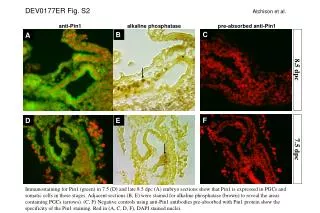
This study investigates the expression of Pin1 in embryonic sections at 7.5 and 8.5 days post-conception (dpc). Using anti-Pin1 antibodies and alkaline phosphatase staining, we reveal that Pin1 is prominently expressed in primordial germ cells (PGCs) and somatic cells during these developmental stages. Adjacent sections were additionally stained for alkaline phosphatase to visualize PGCs, marked with arrows in the imagery. Negative controls confirm the specificity of the Pin1 staining, providing critical insights into its role in early embryonic development.

Immunostaining for Pin1 in Embryo Sections Reveals Expression in PGCs and Somatic Cells
E N D
Presentation Transcript
DEV0177ER Fig. S2Atchison et al. anti-Pin1 alkaline phosphatase pre-absorbed anti-Pin1 C 8.5 dpc B A 7.5 dpc F E D Immunostaining for Pin1 (green) in 7.5 (D) and late 8.5 dpc (A) embryo sections show that Pin1 is expressed in PGCs and somatic cells in those stages. Adjacent sections (B, E) were stained for alkaline phosphatase (brown) to reveal the areas containing PGCs (arrows). (C, F) Negative controls using anti-Pin1 antibodies pre-absorbed with Pin1 protein show the specificity of the Pin1 staining. Red in (A, C, D, F), DAPI stained nuclei.