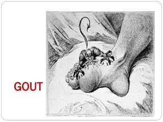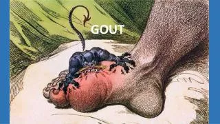
Gout: Causes, Symptoms, and Management
Learn about gout, a disorder caused by uric acid metabolism, leading to joint inflammation and potential damage. Discover its types, risk factors, and associated conditions. Explore its epidemiology, etiology, and pathophysiology.


Gout: Causes, Symptoms, and Management
E N D
Presentation Transcript
Definitions: • Gout is a common disorder of uric acid metabolism that can lead to deposition of monosodium urate (MSU) crystals in soft tissue, recurrent episodes of debilitating joint inflammation, and, if untreated, joint destruction and renal damage. • asymptomatic hyperuricemia is not a disease in the absence of gout
Types gouty arthritis - recurrent attacks of articular and periarticular inflammation • acute gout - acute attacks • intercritical or interval gout - asymptomatic intervals between attacks tophaceous (chronic tophaceous gout)(1, 4, 5) • usually takes many years to progress, not all patients with acute gout will progress to chronic gout • involved joints persistently stiff and swollen • polyarticular involvement may present as subcutaneous nodules that can mimic rheumatoid arthritis • risk factors include early onset of disease, alcohol misuse, persistently elevated serum uric acid levels, and poor compliance with hypouricemic drug therapy • chronic granulomatous inflammatory response surrounding urate crystals “Saturnine gout铅中毒的" - gout associated with lead intoxication(1)
Who is most affected: • male:female ratio 3.6:1 • mean age of onset 40-60 years in men (later in women) and inversely related to serum uric acid levels • rare in premenopausal women(1) • juvenile gout significantly associated with family history of gout and obesity
Incidence/Prevalence: • 0.08% estimated global age-standardized prevalence of gout in 2010. • 2% overall prevalence of self-reported, physician-diagnosed gout in men > 30 years old and women > 50 years old in United States.
Likely risk factors: • hyperuricemia is single most important risk factor for developing gout • risk factors for gout in men • obesity • weight gain • hypertension • diuretic use • alcohol use (dose-dependent) • high levels of sugar sweetened soft drink consumption • high levels of fructose consumption • high levels of meat consumption • high levels of seafood consumption • low levels of dairy product consumption
Possible risk factors: • diuretics, beta-blockers, angiotensin converting enzyme inhibitors, and nonlosartan angiotensin II receptor blockers associated with increased risk for gout in adults with or without hypertension Factors not associated with increased risk: • long-term coffee consumption associated with lower risk of incident gout • calcium channel blockers and losartan associated with reduced risk for gout in adults with hypertension
Associated conditions: • hyperuricemia associated with cardiovascular disease, renal disease, diabetes mellitus, obesity, metabolic syndrome, dyslipidemia, and alcoholism. • most patients with gout have ≥ 1 comorbidity. • gout may be associated with increased risk for type 2 diabetes in males • prevalence of metabolic syndrome higher among patients with gout compared to patients without gout
Causes: • gout caused by inflammation secondary to monosodium urate crystal deposition in joints, peri-articular tissues, or renal tract(1, 4, 5) • hyperuricemia and crystal deposition may be caused by • urate underexcretion (more common than overproduction)(1) • primary hyperuricemia • secondary hyperuricemia • renal impairment /hypertension /drugs -low-dose aspirin.. diuretics.cyclosporine .ethanol/lead nephropathy /hypothyroidism • additional causes of decreased urate excretion may include • ethambutol (Myambutol) /pyrazinamide (Rifater, Tebrazid) /levodopa /niacin (nicotinic acid) /polycystic kidney disease (autosomal dominant polycystic kidney disease) /dehydration /lactic acidosis /hyperparathyroidism /toxemia of pregnancy (hypertensive disorders of pregnancy) /obesity /sarcoidosis
urate overproduction • primary hyperuricemia • hypoxanthine-guanine phosphoribosyltransferase (HPRT)次黄嘌呤鸟嘌呤磷酸核糖转移酶deficiency (Lesch-Nyhan syndrome) • secondary hyperuricemia • excessive dietary purine intake • lympho-/myeloproliferative disorders • severe exfoliative psoriasis次黄嘌呤鸟嘌呤磷酸核糖转移酶 • Drugs: cytotoxic agents /excess ethanol intake /vitamin B12 • uncommon causes of urate overproduction • enzymopathies /chronic hemolysis /rapidly dividing tumors • additional causes of increased urate production may include • excess fructose consumption /obesity /hypertriglyceridemia /warfarin
Pathogenesis: • uric acid is metabolic byproduct of purine catabolism(1, 5) • serum uric acid levels determined by amounts of(2, 3, 4, 5) • purines synthesized and ingested • urate produced from purines • uric acid excreted by kidneys • hyperuricemia has many causes, including combinations of • high purine diet • alcohol use • diuretic therapy • reduced renal clearance • overproduction
Pathogenesis: • hyperuricemia leads to deposition of urate crystals and subsequent inflammation • urate crystals begin precipitating at serum uric acid levels of about 6.8 mg/dL (404 mcmol/L) • abrupt release of urate crystals into joint space may cause acute inflammatory reaction (gouty arthritis) • local factors influencing gout development in presence of hyperuricemia include • trauma (for example, mechanical stress to first metatarsophalangeal joint) • irritation • reduced temperature (for example, helix of ear or foot) • prior joint disease (for example, in Heberden node)
Pathogenesis: • crystals persist in joint after acute attack in intercritical period • often associated with low-grade persistent inflammation • may also persist as microtophi in synovium • may lead to progressive disease (joint damage and erosions) • about 90% of first attacks are monoarticular(4) • usually in lower extremity (midfoot, first metatarsophalangeal joint, ankle, or knee) • additional joints may be affected over time (including upper extremity) • uncommon in axial joints • acute flares may also occur in periarticular structures including(4) • bursae (for example, olecranon and knee) • tendons around ankle
History: Chief concern (CC): • sudden onset of extreme pain, tenderness, and joint inflammation (red, warm, swollen)(1, 4, 5) • may have fever, flu-like malaise(4, 5) History of present illness (HPI): • progression variable(1, 4, 5) • may progress through 4 stages (over many years) if untreated • asymptomatic hyperuricemia • most patients with elevated serum uric acid will not develop gout • 0.5% annual incidence of gout in patients with uric acid level 7-8.9 mg/dL (415-530 mcmol/L) • 4.5% annual incidence of gout in patients with uric acid level ≥ 9 mg/dL (535 mcmol/L)
History: • acute gout • severe pain, erythema, and swelling, often beginning in middle of night or early morning and increasing until peaking within 24-48 hours • usually self-limited with spontaneous resolution in 3-14 days • patients often cannot tolerate socks or weight of bed sheet during acute attack and may be unable to support own weight • about 90% of initial attacks monoarticular • first metatarsophalangeal joint most commonly involved • other frequently involved joints include midfoot, ankles, knees • additional joints may be affected over time (including upper extremity) • uncommon in axial joints • acute bursitis or tenosynovitis may occur in periarticular structures • may resemble cellulitis • skin desquamation may occur over inflamed area
History: • intercriticalor interval gout • intervals between attacks are intercritical periods • subsequent attacks usually longer in duration, involve more joints over time and may not resolve without treatment • crystals usually remain present in periarticular and synovial tissue and may still be present in fluid • chronic tophaceous gout • involved joints persistently stiff and swollen • usually takes many years to progress • frequent recurrent attacks lead to continued accumulation of crystal deposits • intradermal deposits may be white or yellowish, asymptomatic, • polyarticular involvement may present as subcutaneous nodules that can mimic rheumatoid arthritis • rarely, tophi may present as initial manifestation of gout
History: • attacks may have precipitating event, common triggers include(4, 5) • infection • IV contrast media • acidosis • rapid fluctuations in serum uric acid concentrations from • trauma • surgery • psoriasis flares银屑病 • chemotherapy initiation • diuretic therapy • stopping or starting allopurinol • alcohol ingestion
History: Medication history: • hyperuricemia may be caused by(1) • low-dose aspirin /diuretics /cyclosporin /ethanol /cytotoxics /vitamin B12 • uric acid levels may also be increased with • ethambutol (Myambutol) /pyrazinamide (Rifater, Tebrazid) /levodopa/nicotinic acid /didanosine /warfarin Past medical history (PMH): • ask about comorbidities associated with risk for gout • hyperlipidemia /hypertension /metabolic syndrome /chronic kidney disease /obesity /cardiovascular disease /diabetes
History: • urate underexcretion may be caused by • polycystic kidney disease (autosomal dominant polycystic kidney disease) /dehydration /lactic acidosis /hyperparathyroidism /toxemia of pregnancy (hypertensive disorders of pregnancy) sarcoidosis /hypothyroidism(1) Family history (FH): • ask about family history of gout at young age(4) • familial predisposition Social history (SH): • ask about alcohol use • ask about functional impact including limitations in ability to work, meet family responsibilities or enjoy leisure time
Physical: Skin: • acute gout may resemble cellulitis and skin desquamation may occur over inflamed area(5) Extremities: • swollen, red, tender joint during attack • usually unilateral first metatarsophalangeal joint in first attack • most commonly affected joints :great toe /foot /ankle /knee /wrist /finger /elbow • tophi(4, 5) • visible or palpable soft tissue masses • asymptomatic intradermal/subcutaneous nodules or lesions • white or yellowish deposits • overlying skin may be pulled taut
Making the diagnosis: • gold standard is demonstration of urate crystals in synovial fluid analysis or in tophusby polarized light microscopy(1) • American College of Rheumatology (ACR) criteria for classification of acute gouty arthritis • presence of characteristic urate crystals in joint fluid, OR • tophus proven to contain urate crystals by chemical means or polarized light microscopy, OR • 6 of the following 12 criteria (not a definitive diagnosis) • more than 1 attack of acute arthritis • maximal inflammation developed within 1 day • attack of monarticular arthritis
Making the diagnosis: • joint redness observed • first metatarsophalangeal joint painful or swollen • unilateral attack involving first metatarsophalangeal joint • unilateral attack involving tarsal joint • suspected tophus • hyperuricemia • asymmetric swelling within a joint (radiograph) • subcortical cysts without erosions (radiograph) • negative culture of joint fluid for microorganisms during attack of joint inflammation • presumptive diagnosis can be made based on following • presence of hyperuricemia
Making the diagnosis: • careful patient and family history including questions regarding • comorbidities (for example, hypertriglyceridemia, diabetes, coronary heart disease, hypertension, metabolic syndrome) • previous similar episodes of acute joint pain and swelling in absence of trauma • identification of current medications that may be associated with hyperuricemia • physical exam including • joints • extensor surfaces of forearms and feet • common sites for tophi (for example, ear, knee, olecranon bursa) • other clinical features • duration of joint pain • fever (low-grade or high)
Making the diagnosis: • careful patient and family history including questions regarding • comorbidities (for example, hypertriglyceridemia, diabetes, coronary heart disease, hypertension, metabolic syndrome) • previous similar episodes of acute joint pain and swelling in absence of trauma • identification of current medications that may be associated with hyperuricemia • physical exam including • joints • extensor surfaces of forearms and feet • common sites for tophi (for example, ear, knee, olecranon bursa) • other clinical features • duration of joint pain • fever (low-grade or high)
Differential diagnosis • calcium pyrophosphate dihydrate (CPPD) deposition disease (pseudogout)(5) • gram-negative stain • rhomboid长菱形shaped crystals with weak positive birefringence双折射性in synovial fluid • soft tissue swelling or chondrocalcinosis on x-ray • septic arthritis(5) • knee most commonly involved • joint effusions on x-ray • bacterial cellulitis(cutaneous erythema may extend beyond involved joint)
Differential diagnosis • rheumatoid arthritis (RA)(4) • crystal deposition can cause chronic polyarthritis and mimic RA • elderly patients may develop rheumatoid factor positivity • tophaceous gout may be distinguished from rheumatoid arthritis by • presence of urate crystals in aspirate of tophus or synovial fluid • radiographic exam • psoriatic银屑病arthritis(4) • erosive osteoarthritis
Testing overview: • synovial fluid analysis and culture(5) • complete blood count, blood urea nitrogen, creatinine • serum uric acid • imaging studies allow visualization of affected joint and may include • x-ray • ultrasound • computed tomography • blood culture if suspecting septic arthritis
Blood tests: • serum uric acid level • limited usefulness during acute attack(4, 5) • uric acid level may be normal during acute gout attack • uric acid > 6.8 mg/dL (404 mcmol/L) sufficient to precipitate crystals, although some laboratories may list higher upper limits of normal(4) • serum urate levels helpful in monitoring effects of antihyperuricemic therapy(3) • leukocytosis may occur in acute gout(5)
Urine studies: • 24-hour urine uric acid measurement not routinely performed(3, 5) • useful for patients being considered for uricosuric therapy or when identifying and excluding urate overproducers • urinary uric acid excretion > 800-1,000 mg/24 hours suggests urate overproduction and increased risk of uric acid kidney stones
Imaging studies: • x-ray(4, 5) • asymmetric swelling with acute gout • prominent, proliferative bony reaction • bone destruction away from joint may be caused by tophi • characteristic "overhanging edge" or "rat bite" of proliferating bone surrounding erosion may be present • gout less likely to cause joint space narrowing than psoriatic arthritis or rheumatoid arthritis
Imaging studies: • ultrasound • features for acute gout nonspecific and include • periarticular soft-tissue edema • hypervascularity within and around joint • features for chronic gout • hyperechoic, irregular band over superficial margin or articular cartilage (double contour sign) • tophi appearing as hypoechoic to hyperechoic inhomogeneous areas surrounded by small anechoic rim • less specific features may be observed using Power Doppler • bone erosion /joint effusions /synovial hypertrophy /hypervascularity • for diagnosing gout, x-ray may be more specific and ultrasound may be more sensitive .
Imaging studies: • computed tomography (CT) • may detect changes of early disease that are not visible on plain radiography(6) • CT of affected joint may be useful for • visualizing tophi, especially intra-articular tophi (mean Hounsfield units of tophi usually 170 on CT) • visualizing bone erosion • guiding needle aspiration • assessing complications • CT measurement of tophus has good correlation with physical measurement
Imaging studies: • dual-energy CT may be useful for diagnosis of non-tophaceous gout (level 2 [mid-level] evidence) • for diagnosis of gout, dual-energy CT had • sensitivity 90% • specificity 83% • positive predictive value 84% • negative predictive value 90% • dual-energy CT may be highly specific for detection of gout • CT reported to provide more specific images of tophaceous gout than x-ray, ultrasound, or magnetic resonance imaging
Biopsy and pathology: • tophus is pathognomonic • biopsy of tophus shows chronic foreign body granulomatous inflammation surrounding monosodium urate crystals(4) • fine needle aspiration of gouty tophus • small whitish material may be visible macroscopically • microscopic appearance • aggregates of crystalline material • occasional histiocytes • multinucleate giant cells less common • slender, rod-shaped crystals with pointed ends in smear background • crystals strongly (negatively) birefringent • crystals may also appear as aggregates of dense, amorphous material that stain dark grayish with Giemsa-based stains
Other diagnostic testing: • synovial fluid analysis • characteristics of monosodium urate crystals • needle shaped • about 2-20 mm in length • strong negative birefringence under polarized light • appear yellow when parallel to axis of slow vibration of compensator and appear blue when perpendicular to axis • usually intracellular during acute attacks and intercritical periods • mostly extracellular and free in synovial fluid in chronic gout • in presence of fever and elevated white blood cell count, aspiration and analysis of synovial fluid must be performed to exclude septic arthritis, either alone or coexisting with gout
Treatment overview: • for acute attack • rest and elevate affected joints • ice packs • nonsteroidal antiinflammatory drugs (NSAIDs) often drug of choice and different NSAIDs appear equally effective in optimum doses • colchicine (1.2 mg orally then 0.6 mg 1 hour later) appears effective but slower to work than NSAID • alternative drugs for aborting acute attack include • corticosteroids /corticotropin /canakinumab (Ilaris)人抗白介素-1β单克隆抗体
Treatment overview: • for prevention of recurrent attacks • urate-lowering therapy recommended if ≥ 2 attacks per year .tophi .uric acid stone or reduced kidney function . • target serum uric acid level ≤ 6 mg/dL (360 mcmol/L) but some patients may require level < 5 mg/dL (300 mcmol/L) to control symptoms • first-line options for urate-lowering therapy are • allopurinol 50-100 mg/day orally, increased up to maximum 800-900 mg/day (ACR Evidence A; BSR Grade B; EULAR Level Ib) • febuxostat (Uloric) 40-80 mg orally once daily (ACR Evidence A)
Treatment overview: • second-line options for urate-lowering therapy are uricosuric drugs (such as probenecid, sulfinpyrazone, or benzbromarone) • uricolytic enzymes, such as pegloticase (Krystexxa)聚乙二醇尿酸酶, may be effective for severe gout refractory to conventional urate-lowering therapy. • anti-inflammatory prophylaxis (with colchicine 0.5-0.6 mg once or twice daily, NSAID, or corticosteroid) recommended for all gout patients when urate-lowering therapy is started (ACR Evidence A) and continued for at least 6 months (ACR Evidence A) and if any clinical disease activity or elevated serum uric acid level • restrict intake of high purine foods, red meat, and alcohol
Complications: • short-term disability in most patients with acute gouty attack • gout (and hyperuricemia) may be associated with increased risk of coronary artery disease • renal disease • hyperuricemia may be associated with renal failure • forms of hyperuricemia-induced renal disease include • uric acid nephrolithiasis (kidney stones) • acute uric acid nephropathy (associated with chemotherapy and tumor lysis syndrome) • chronic kidney disease resulting from urate crystal deposition • complications of tophaceous gout • joint erosion or destruction(1) /carpal tunnel syndrome caused by tophaceous gout in case report
Prognosis: • first acute attack usually subsides in 3-14 days(4) • subsequent attacks usually last longer and may involve more joints(4) • risk of recurrence after initial attack(4) • about 60% recurrence within 1 year • about 78% within 2 years • about 84% within 3 years • > 90% will have recurrence at 10 years • serum uric acid levels > 6 mg/dL (360 mcmol/L) associated with increased risk for recurrent gout attacks • development of tophi associated with(1) • early onset of disease • alcohol misuse • persistently elevated uric acid levels • poor compliance with hypouricemic drug therapy • diuretic use in renal insufficiency and heart failure (especially in women) • cyclosporin use with organ transplant
Prevention: • modifiable risk factors include(5) • high-purine diet • alcohol use • obesity • diuretic therapy • consumption of dairy products may be protective(5) • long-term coffee consumption associated with lower risk of incident gout (level 2 [mid-level] evidence) • higher vitamin C intake associated with lower risk of gout (level 2 [mid-level] evidence)
广济医院(现“浙医二院”)首任院长梅腾更先生与小患者互相鞠躬致敬广济医院(现“浙医二院”)首任院长梅腾更先生与小患者互相鞠躬致敬 感谢!