Download
1 / 61
610 likes | 677 Vues
Learn about the intricate processes of fluid secretion, absorption, and enzyme activity in the small intestine, crucial for breaking down food and absorbing nutrients effectively. Understand the mechanisms behind mixing and propulsive contractions that aid digestion.

E N D
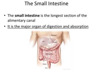
Digestion in the small intestine • Approximately 1500 ml of fluid is secreted by the walls of the small intestine from the blood into the lumen each day. • One of the reasons for water movement into the lumen (secretion) is that the intestinal epithelium at the base of the villi secretes a number of mineral ions, notably sodium, chloride, and bicarbonate ions into the lumen, and water follows by osmosis.
Digestion in the small intestine • Normally, all of the fluid secreted by the small intestine is absorbed back into the blood →a large net absorption of water. • Absorption is achieved by the transport of ions, primarily sodium, from the intestinal lumen into the blood, with water following by osmosis.
Digestion in the small intestine Brunner’s glandssecrete large amounts of alkaline mucus in response to (1) tactile or irritating stimuli on the duodenal mucosa; (2) vagal stimulation, which causes increased Brunner’s glands secretion concurrently with increase in stomach secretion; and (3) gastrointestinal hormones, especially secretin
Digestion in the small intestine Crypts of Lieberkühnlie between the intestinal villi. The surfaces of the crypts and the villi are covered by an epithelium composed of two types of cells: (1) a moderate number of goblet cells, which secrete mucus that lubricates and protects the intestinal surfaces, (2) a large number of enterocytes, which, in the crypts, secrete large quantities of water and electrolytes.
Digestion in the small intestine • The mechanism that controls the marked secretion of watery fluid by the crypts of Lieberkühn is not known. It is believed to involve at least two active secretory processes: (1) active secretion of Cl- into the crypts and (2) active secretion of bicarbonate ions causes electrical drag as well of positively charged sodium ions through the membrane and into the secreted fluid. Finally, all these ions together cause osmotic movement of water.
Digestion in the small intestine The enterocytes of the mucosa, especially those that cover the villi, contain digestive enzymes that digest specific food substances while they are being absorbed through the epithelium. (1) several peptidases for splitting small peptides into amino acids, (2) four enzymes—sucrase, maltase, isomaltase, and lactase—for splitting disaccharides into monosaccharides, and (3) small amounts of intestinal lipase for splitting neutral fats
Regulation of small intestine secretion • By far the most important means for regulating small intestine secretion are local enteric nervous reflexes, especially reflexes initiated by tactile or irritative stimuli from the chyme in the intestines.
Movements of the small intestine The movements of the small intestine, can be divided into • mixing contractions and • propulsive contractions
Mixing (Segmentation)contractions • When a portion of the small intestine becomes distended with chyme, stretching of the intestinal wall elicits localized concentric contractions spaced at intervals along the intestine and lasting a fraction of a minute. • Each contracting segment is only a few centimeters long and the contraction lasts a few seconds. The chyme in the lumen of a contracting segment is forced both up and down the intestine.
Mixing (Segmentation ) contractions • This rhythmical contraction and relaxation of the intestine, segmentation, produces a continuous division and subdivision of the intestinal contents, thoroughly mixing the chyme in the lumen and bringing it into contact with the intestinal wall. • That is, they divide the intestine into spaced segments that have the appearance of a chain of sausages.
Mixing(Segmentation)contractions • The maximum frequency of the segmentation contractions in the small intestine is determined by the frequency of electrical slow waves in the intestinal wall, which is the basic electrical rhythm. • Because this frequency normally is not over 12 per minute in the duodenum and proximal jejunum; in the terminal ileum, the maximum frequency is usually 8 to 9 contractions per minute
Propulsive movements peristalsis in the small intestine • Chyme is propelledthrough the small intestine by peristaltic waves. These can occur in any part of the small intestine and they move toward the anus at a velocity of 0.5 to 2.0 cm/sec, faster in the proximal intestine and slower in the terminal intestine. • They normally are very weak andusually die out after traveling only 3 to 5 centimeters, very rarely farther than 10 centimeters, so that forward movement of the chyme is very slow.
Propulsive movements peristalsis in the small intestine • Peristaltic activity of the small intestine is greatly increased after a meal • is caused partly by the beginning entry of chyme into the duodenum causing stretch of the duodenal wall, but also by a so-called gastroentericreflex that is initiated by distention of the stomach and conducted principally through the myenteric plexus from the stomach down along the wall of the small intestine
Propulsive movements peristalsis in the small intestine • In addition to the nervous signals that may affect small intestinal peristalsis, several hormonal factors also affect peristalsis: gastrin, CCK, insulin, motilin, and serotonin, all of which enhance intestinal motility and are secreted during various phases of food processing. • Conversely, secretin and glucagoninhibit small intestinal motility.
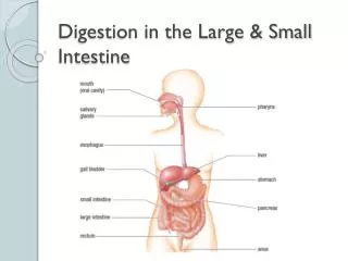
Large intestine • The colonconsists of three relatively straight segments—the ascending, transverse and descending portions. • The terminal portion of the descending colon is S-shaped, forming the sigmoid colon, which empties into a relatively straight segment of the large intestine, the rectum, which ends at the anus.
Large intestine • The secretions of the large intestine are scanty, lack digestive enzymes and consist mostly of mucus and fluid containing bicarbonate and potassium ions. • The primary function of the large intestine is to store and concentrate fecal material before defecation.
Large intestine • The primary absorptive process is the active transport of Na+ from lumen to blood, with the accompanying osmotic absorption of water. If fecal material remains in the large intestine for a long time, almost all the water is absorbed, leaving behind hard fecal pellets. There is normally a net movement • of K+ from blood into the largeintestine lumen • bicarbonate ions into the lumen, and loss of this bicarbonate (a base) in patients with prolonged diarrhea can cause the blood to become acidic.
Large intestine • The large intestine also absorbs some of the products formed by the bacteria inhabiting this region. • Undigested polysaccharides (fiber) are metabolized to short-chain fatty acids by bacteria in the large intestine and absorbed by passive diffusion. • The bicarbonate secreted by the large intestine helps to neutralize the increased acidity resulting from the formation of these fatty acids
Large intestine • The mucosa has many crypts ofLieberkühn; however, unlike the small intestine, thereare no villi. • The epithelial cells contain almost no enzymes; they consist mainly of mucous cellsthat secrete only mucus. • This mucus contains moderate amounts of bicarbonate ions. • The rate of secretion of mucus is regulated principally by direct, tactile stimulation and by local nervous reflexes.
Large intestine • Stimulation of the pelvic nerves from the spinal cord, which carry parasympathetic innervation to the distal one half to two thirds of the large intestine, also can cause marked increase in mucus secretion.
Large intestine Movements of the colon The principal functions of the colon are • absorption of water and electrolytes from the chyme to form solid feces and • storage of fecal matter until it can be expelled. The proximal half of the colon, is concerned principally with absorption and the distal half with storage.
Large intestine • Because intense colon wall movements are not required for these functions, the movements of the colon are normally very sluggish. • the movements still have characteristics similar to those of the small intestine and can be divided once again into mixing movements and propulsive movements.
Large intestine • In the same manner that segmentation movements occur in the small intestine, large circular constrictions occur in the large intestine. • At each of these constrictions, about 2.5 centimeters of the circular muscle contracts, sometimes constricting the lumen of the colon almost to occlusion.
Large intestine • At the same time, the longitudinal muscle of the colon, which is aggregated into three longitudinal strips called the teniae coli, contracts. • combined contractions of the circular and longitudinal strips of muscle cause the unstimulated portion of the large intestine to bulge outward into baglike sacs called haustrations. • Each haustration usually reaches peak intensity in about 30 sec and then disappears during the next 60 seconds.
Large intestine • Contractions of the circular smooth muscle in the large intestine produce a segmentation motion with a rhythm considerably slower (one every 30 min) than that in the small intestine. • Because of the slow propulsion of the large intestine contents, material entering the large intestine from the small intestine remains for about 18 to 24 h. This provides time for bacteria to grow and multiply.
Large intestine • Three to four times a day, generally following a meal, a wave of intense contraction, known as a mass movement, spreads rapidly over the transverse segment of the large intestine toward the rectum. • Unlike a peristaltic wave, in which the smooth muscle at each point relaxes after the wave of contraction has passed, the smooth muscle of the large intestine remains contracted for some time after a mass movement.
Digestion and absorption in the gastrointestinal tract Hydrolysis of carbohydrates Almost all the carbohydrates of the diet are eitherlarge polysaccharides or disaccharides . When carbohydrates are digested, they are converted into monosaccharides. Specific enzymes in the digestive juices of the gastrointestinal tract return the hydrogen and hydroxyl ions from water to the polysaccharides and thereby separate the monosaccharides from each other.
Digestion and absorption in the gastrointestinal tract Hydrolysis of fats • Almost the entire fat portion of the diet consists of triglycerides (neutral fats)=combinations of three fatty acid molecules condensed with a single glycerol molecule). During condensation, three molecules of water are removed. • Digestion of the triglycerides consists of the reverse process: the fatdigesting enzymes return three molecules of water to the triglyceride molecule and thereby split the fatty acid molecules away from the glycerol.
Digestion and absorption in the gastrointestinal tract Hydrolysis of proteins • P =multiple amino acids that are bound together by peptide linkages. At each linkage, a hydroxyl ion has been removed from one amino acid and a hydrogen ion has been removed from the succeeding one; thus, the successive amino acids in the protein chain are bound together by condensation; digestion occurs by the reverse effect. • The proteolytic enzymes return hydrogen and hydroxyl ions from water molecules to the protein molecules to split them into their constituent AA.
Digestion and absorption in the gastrointestinal tract • The total quantity of fluid that must be absorbed each day by the intestines is equal to the ingested fluid (about 1.5 liters) plus that secreted in the various gastrointestinal secretions (about 7 l)=total of 8 to 9 liters. • 1.5 liters of this is absorbed in the small intestine, leaving only 1.5 liters to pass through the ileocecal valve into the colon • The stomach is a poor absorptive area!
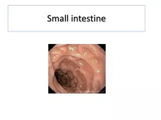
Digestion and absorption in the gastrointestinal tract • The absorptive surface of the small intestinal mucosa, shows folds called valvulae conniventes (or folds of Kerckring) • These folds extend circularly most of the way around the intestine and are especially well developed in the duodenum and jejunum • on the epithelial surface of the small intestine all the way down to the ileocecal valve are literally millions of small villi.
Digestion and absorption in the gastrointestinal tract • Thus, the combination of the valvulae conniventes, the villi, and the microvilli increases the total absorptive area of the mucosa perhaps 1000-fold, making a tremendous total area of 250 or more square meters for the entire small intestine.
Absorption of sodium • 20 to 30 grams of sodiumare secreted in the intestinal secretions each day. In addition, the average person eats 5 to 8 grams of sodium each day. • Therefore, to prevent net loss of sodium into the feces, the intestines must absorb 25 to 35 grams of sodium each day, which is equal to aboutone seventh of all the sodium present in the body.
Chloride ion absorption • In the upper part of the small intestine, chloride ion absorption is rapid and occurs mainly by diffusion— that is, absorption of sodium ions through the epithelium creates electronegativity in the chyme and electropositivity in the paracellular spaces between the epithelial cells. • Then chloride ions move along this electrical gradient to “follow” the sodium ions.
Absorption of the bicarbonate • an indirect way: when sodium ions are absorbed, moderateamounts of hydrogen ions are secreted into the lumenof the gut in exchange for some of the sodium. • thesehydrogen ions in turn combine with the bicarbonateions to form H2CO3→ form H2O and CO2. • H2O remains as part of the chyme in the gut, • CO2 is absorbed into the blood and subsequently expired through the lungs.
Absorption of the bicarbonate • The epithelial cells on the surfaces of the villi in the ileum as well as on all surfaces of the large intestine have a special capability of secreting bicarbonate ions in exchange for absorption of chloride ions. • This is important because it provides alkaline bicarbonateions that neutralize acid products formed by bacteria in the large intestine.
Ions absorption • Calcium ionsare actively absorbed into the blood especially from the duodenum • absorption is very exactly controlled to supply exactly the daily need of the body for Ca. • controlling calcium absorption is parathyroid hormone secreted by the parathyroid glands, and another is vitamin D. • parathyroid hormone activates vitamin D and the activated vitamin D in turn greatly enhances calcium absorption
Ions absorption • Iron ions are also actively absorbed from the small intestine. • Potassium, magnesium, phosphate, and probably still other ions can also be actively absorbed through the intestinal mucosa. In general, the monovalent ions are absorbed with ease and in great quantities. • Conversely, bivalent ions are normally absorbed in only small amounts; for example, maximum absorption of calcium ions is only 1/50 as great as the normal absorption of sodium ions.
Absorption of water • Entirely by diffusion (obeys the usual laws of osmosis). • When the chyme is dilute enough, water is absorbed through the intestinal mucosa into the blood of the villi almost entirely by osmosis. • Conversely, water can also be transported in the opposite direction—from plasma into the chyme. This occurs especially when hyperosmotic solutions are discharged from the stomach into the duodenum.
Carbohydrates absorption • Essentially all the carbohydrates in the food are absorbed in the form of monosaccharides; only a small fraction are absorbed as disaccharides and almost none as larger carbohydrate compounds. • By far the most abundant of the absorbed monosaccharides is glucose
Carbohydrates absorption • In the absence of sodium transport through the intestinal membrane, virtually no glucose can be absorbed. • The reason is that glucose absorption occurs in a cotransport mode with active transport of sodium
Carbohydrates absorption • There are two stages in the transport of sodium through the intestinal membrane. • active transport of sodium ions through the basolateral membranes of the intestinal epithelial cells into the blood, thereby depleting sodium inside the epithelial cells • decrease of sodium inside the cells causes sodium from the intestinal lumen to move through the brush border of the epithelial cells to the cell interiors by a process of facilitated diffusion.
Carbohydrates absorption • intestinal glucose also combines with the same transport protein • the low concentration of sodium inside the cell literally “drags” sodium to the interior of the cell and along with it the glucose at the same time. • inside the epithelial cell, other transport proteins and enzymes cause facilitated diffusion of the glucose through the cell’s basolateral membrane into the paracellular space and from there into the blood.