Download
1 / 46
620 likes | 1.85k Vues
Golgi complex, secretion and protein transport. Biology I – Faculty of Pharmacy Dr. Eszter Lajkó Department of Genetics, Cell- and Immunobiology 03.10.2016. Protein targeting and sorting Transport of proteins from the synthesis site to their destinations.

E N D
Golgi complex, secretion and protein transport Biology I – Faculty of Pharmacy Dr. Eszter Lajkó Department of Genetics, Cell- and Immunobiology 03.10.2016
Protein targeting and sorting Transport of proteinsfromthesynthesis site totheirdestinations
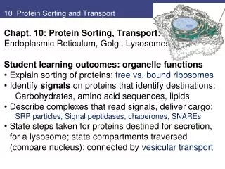
Key components of the protein transport • Sortingsignal • Receptors: recognizesortingsignal and guideproteinstotheirappropriatedestination • Way of protein transfer • Gatedtransportthroughthenuclearpore • Translocationacrossthemembrane (transmembrane protein traslocator = translocon) • Vesiculartransport • Energy
Protein sorting Co-translational transmembrane transport Protein synthesis in cytoplasm (free ribosome) Endoplasmic reticulum (membrane-bounded ribosome) (hydrophobic aa at N-term) Gated transport Vesicular transport Post-translational transmembrane transport Cytoplasm (no signal) Golgi Nucleus (NLS) Late endosome Secretory vesicle Mitochondria (N-term. positively charged aa) Lysosome Early endosome Peroxisomes (3 aa at C-term) Plasma membrane
Vesiculartransport • transport between membrane-enclosed compartments • transport of macromolecules (soluble and membrane-bound) from the donor compartment to the target compartment Further information about the vesicular transport in 6th week lecture
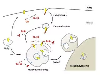
Main vesiculartransportpathways Inward transport Endocytotic pathway Outward transport Secretory pathway
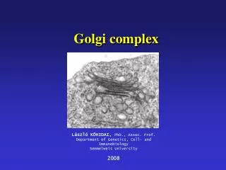
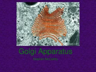
Golgi apparatus "internalreticularapparatus" Camillo Golgi (1843-1926) Nobel prize 1906 Metal impregnation
Structure of Golgi apparatus • consist of saccules(cisternae), tubules and vesicles • structural-functional unit: dictyosome(4-6 saccules) • structure is polarized into sub-compartments entry Endoplasmic reticulum Cis Golgi network (CGN) Cis Golgi (CG) Medial Golgi (MG) Trans Golgi (TG) Trans Golgi network (TGN) exit Plasma membrane Lysosome
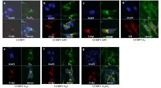
Position Nucleus Microtubule Golgi
Role of microtubules in maintance of Golgi structure IntactmicrotubulesDisintegration of microtubules Golgi- green Microtubules- red 2002 by Bruce Alberts, Alexander Johnson, Julian Lewis, Martin Raff, Keith Roberts, and Peter Walter.
Visualisation of Golgi withdifferenttechniques Silver impregnation for LM TEM 1. Golgi dyctiosomes Multiphoton fluorescence image
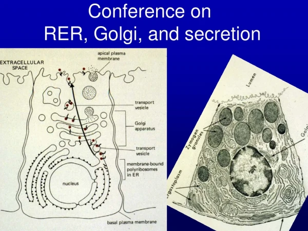
Main functions Sructural and functional polarization Cis face: arrival side post-translational modifications of proteins (and lipids) sorting formation of transport vesicles transport trans face: departure side
Vesiculartubularcluster • continually generated from ER-derived vesicles • separate compartment from ER and Golgi • transport container • moving along the microtubules to the Golgi
Cis Golgi Network – Entry side • collection of fused vesicular tubular clusters • the proteins and lipids arrive from the ER in vesicles move back to the ER • ER-resident protein • proteins of Golgi • targeting to the cell surface or another compartments move to the cis Golgi cisterna • they are N-glycosylated • no sorting in the ER
Glycosylation N-glycosylation O-glycosylation • Adding oligosaccharide chain to protein (lipids) • Made by glycosyl transferases • N-glycosylation (to Asn) is made in ER and modified in ER and Golgi(longer chains) • O-glycosylation (to Ser, Thr) is made in Golgi (shorter chains) soluble glycoportein O-glycosylation Glycolipid N-glycosylation Membrane glycoportein
N-glycosylation • Preformed oligosaccharide chain is transfered to asparagine side chains of a polypeptide in ER • Processing of oligosaccharide chain starts in ER and continues in Golgi cytoplasm Asparagine side chain ER lumen Golgi: further processing of N-linked olgosaccharide chain
Modificationof N-oligosaccharide chains of proteins • phosphorylationof the mannoses of thelysosomal proteins • cisGolgi network • no change (high mannose) • cis Golgi • change of mannoses to other monosaccharides • medial and trans Golgi • each cisternae containing a characteristic mixture of enzymes involved in processing of N-linked oligosaccharides • enzymes are all membrane bound • processing depends on the position of oligosaccharides in the protein
Sorting and modification of lysosomal enzymes • Mannose-6-phosphate (M-6-P) signal • basedontherecognition of lysosomalhydrolases • recognitionof the“signal patches” is required • main workingenzyme: GlcNAc-phosphotransferase
Significance of M-6-P labelling Lysosomal enzyme Lysosome Mannose-6-phosphate M-6-P • Lysosomalenzymeswith M-6-P signalare • not modified further in Golgi • recognisedbyreceptors in trans Golgi network • transportedtothelysosome Golgi
Main types of N-oligosaccharidechains -Asn -Asn -Asn
Steps of processing and subsequent sugar addition is rigidly ordered, but the complex oligosaccharides can be heterogeneous medial Golgi cis Golgi medial Golgi trans Golgi negatively charged trans Golgi
N- and O-linkedglycosylation Threonine, Serine
O-linkedglycosylation -Ser/Thr -Ser/Thr • takes place mainly in the medial- and trans Golgi • shorter oligosaccharide chains • adding monosaccharides one by one to Ser/Thr • each step requires different enzymes
Significance of glycosylation • folding • sorting • protection against proteolytic enzymes • makes proteins hydrophylic • cell adhesion (leukocytes and endothel – cell adhesion molecules) • antigenity (A,B,O blood groups) • glycocalyx (external coat)
Proteoglycansynthesis Basic structure of proteoglycans Serine linker: tetrasaccharide Glycosaminoglycan (GAG) core protein • Proteins with polisacharide side chains (long, unbranched) • Found in plasmambrane and extracellular matrix • Major components: glycosaminoglycan (GAG) • composed of repated disaccharides • containing sulphate group negatively charged • e.g. hyaluronic acid, chondroitin sulphate, dermatan sulphate, heparan sulphate
Proteoglycans N and O oligosaccharides are not shown
Modifications of proteins and lipids in Golgi • Glycosylation • modificationof oligosaccharidechainsofproteins, • bindingof newlysynthesizedoligo-orpolysaccharidechains • M-6-P group (lysosomalproteins) • no change (highmannose) • changeof mannosestoothermonosaccharides • O-glycosylation • proteoglycanssynthesis • addingof -SO4group • carbohydrates: GAG, proteoglycans • aminoacid: Tyrresidue of proteins • synthesisof lipids • glycolipids • sphingomyelin • proteolysis chondroithine sulphate
Synthesis of membrane lipids Golgi Sphingomyelin ER Oligosaccharide Monosaccharide Cerebroside Gangliosode Clycolipid
A,B,0 bloodgroupantigens (glycolipids = ganglioside) ceramide sugar residues
Golgi is a major protein sorting • back toER • ER residentproteins (soluble) – KDEL signal • membraneproteins – KKXX signal • cargoreceptors • proteinsneededforvesiculeformation and fusion • retaining Golgi proteins • aggregation of Golgi proteins • tolysosome • M-6-P signal • toplasmamembrane and ECM • tocellexterior(forsecretion) cis Golgi network and from all cisternae in all cisternae trans Golgi network
BidirectionaltransportbetweenER and Golgi Vesicular tubular cluster Secretory protein ER-resident protein receptor (KDEL receptor) ER-resident protein with KDEL signal sequence
Transportthroughthe Golgi apparatus Cisternal maturation model Vesicle transport model • Cisternae mature from early to late by acquiring and then losing specific Golgi-resident proteins • Cisternae moves through the dictyosome with cargo in its lumen • Retrograde transport of Golgi enzymes • Cisternae remain at the same place with characteristic set of Golgi proteins • Cargo protein are moved forward by transport vesicle • Retrograd pathway of vesicles return the escaped proteins to the previous cisternae
Golgi residentproteins - enzyme-content of thedifferentcompartments - medial Golgi mannosidase II cis Golgi mannosidase I trans Golgi nucleoside diphosphatase Unstained Osmium impregnation cis face trans face
Bidirectionaltransport of proteins • Forward – anterograd transport • lysosomal enzymes • secretory proteins • extracellular matrix and plasma membrane components • Golgi-resident proteins • Backward – retrograd transport • proteins of the ERrecycled • membrane-bound or soluble • retention signal is required • protein of each cisterna • M-6-P receptor from the lysosomes • membrane components of transport vesicles from the plasma membrane
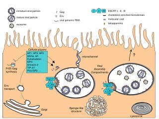
Main transport pathways from TGN Proteins are seggregated into different transport packages and dispatched exocytosis transport from the TGN to the cell exterior (fusion of transport vesicles with plasma membrane) secretory vesicles • secretionsynthesis, modification and release (exocytosis) of different compounds (e.g. proteins, lipids) • centralorganelles – ER and Golgi • types: • consitutivesecretion • regulatedsecretion lysosome endosomal-lysosomal compartment
Constitutive secretion (exocytosis) • defaultpathway • presents in allcells • continuously • non-selective • no accumulation of vesicles • ECM proteins, membranelipidsand proteins signal
Regulated secretion (exocytosis) • typicalforglandularcellsand neurons • signal is neededfortheexocytosis • accumulation of vesicles • hormones, eurotransmitters, digestiveenzymes signal
Modifications of secretory vesicles • selective aggregation - TGN • further modifications and sorting • inactive precursor - active enzyme or hormone • (e.g. preproinsulin - proinsulin - insulin) • concentration - loss of water • hydratation - e.g. proteoglygans • uptake some cytoplasmatic substances e.g. histamine
Immature secretory vesicle contains missorted proteins Removal of missorted proteins Acidification and condensation
Proteolysis of proteins in secretory vesicles proteolysis pro-hormone inactive Synthesis ofpre-pro-hormone inactive active hormone hormone active Secretory vesicle with pro-hormone inactive Glycolistaion of pro-hormone inactive