Costochondritis
Costochondritis. Developed for OUCOM CORE By: Sheri Hull, D.O. Edited by Wayne Feister, D.O. and the CORE Osteopathic Principles and Practices Committee Series C – Session #7. Definition. Inflammation of the junction of the upper ribs and the costal cartilage

Costochondritis
E N D
Presentation Transcript
Costochondritis Developed for OUCOM CORE By: Sheri Hull, D.O. Edited by Wayne Feister, D.O. and the CORE Osteopathic Principles and Practices Committee Series C – Session #7
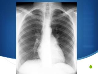
Definition • Inflammation of the junction of the upper ribs and the costal cartilage • Localized chest pain aggravated by coughing, sneezing or deep breathing • aka: Anterior Chest Wall Syndrome
Causes of costochondritis are not known and may involve several factors. Possible causes include heredity (genetic predisposition), viruses, and trauma (injury). Costochondritis can be an independent condition by itself or sometimes be a feature of a more widespread disorder. Examples of illnesses that can feature costochondritis include fibromyalgia, psoriatic arthritis, ankylosing spondylitis, reactive arthritis and inflammatory bowel disease, i.e. ulcerative colitis and Crohn’s disease. Causes of Costochondritis
Anatomy Netter, F. Atlas of Human Anatomy. 2nd Edition. 1997. East Hanover: Novartis
Anatomy Netter, F. Atlas of Human Anatomy. 2nd Edition. 1997. East Hanover: Novartis
Symptoms • Chest Pain • Very common in children and adults • Sharp chest pain with/without radiation to back • 4th, 5th, 6th ribs most common • Reproducible by palpating the costochondral joint
Differential Diagnosis • Cardiac Conditions • Tietze Syndrome • - An inflammation of the costochondral cartilages of the upper front of the chest. Blood testing (Sedimentation Rate or C-Reactive Protein Rate) can show signs of inflammation in patients with Tietze syndrome, whereas patients with costochondritis alone typically have normal tests for inflammation.
Diagnosis - Costochondritis • Motion may be restricted – may be necessary to correct rib motion restrictions to make accurate diagnosis • Based on painful palpitation of the costochondral junction • Also dependent on exclusion of other causes (cardia, infections, etc.)
Treatment • Direct Rib Release – • Remember these people are in pain! • Anti-inflammatory medication and moist heat may also offer relief
Treatment Direct Rib Release Speece, C., et. al. Ligamentous Articular Strain: Osteopathic Manipulative Techniques for the Body. 2001. Seattle:Eastland Press.
Treatment Direct Rib Release • The physician presses upward with the bottom hand while releasing pressure from the top hand. • The physician holds this position for several seconds, after which the bottom hand releases pressure and the top hand exerts downward pressure. Nicholas: Atlas of Osteopathic Techniques
Treatment Muscle Energy • During exhalation the physician’s right hand exaggerates the exhalation motion of the dysfunctional rib. • The patient inhales again (black arrow) as the physician’s right hand resists (white arrow) the inhalation motion of the dysfunctional rib. Nicholas: Atlas of Osteopathic Techniques
Treatment Muscle Energy The patient exhales, and the physician exaggerates the exhalation motion (white arrow) of the dysfunctional rib. Nicholas: Atlas of Osteopathic Techniques
Treatment HVLA • The physician slightly rolls the patient toward the physician by gently pulling the left posterior shoulder girdle forward. • The physician places the thenar eminence of the right hand posterior to the angle of the dysfunctional rib. • The patient is rolled back over the physician’s hand, and the surface created by the patient’s crossed arms rests against the physician’s chest or abdomen. Nicholas: Atlas of Osteopathic Techniques
Treatment HVLA • Pressure is directed through the patient’s chest wall, localizing at the thenar eminence. • The patient inhales and exhales, and at end exhalation a thrust impulse (white arrows) is delivered through the patient’s chest wall slightly cephalad to the thenar eminence. Nicholas: Atlas of Osteopathic Techniques
References • Downing, CH. Osteopathic Principles in Disease. 1935. AAO. • DiGiovanna, E. & Schiowitz, S. An Osteopathic Approach to Diagnosis and Treatment. 1997. Philadelphia: Lippincott, Williams, and Wilkins. • Kimberly, PE. Outline of Osteopathic Manipulative Procedures: The Kimberly Manual, Millennium Edition. 2000. Marceline: AOA. • Foundations for Osteopathic Medicine. 1997. Baltimore: Williams and Wilkins. • Nicholas, AS. Atlas of Osteopathic Techniques. 2008 Lippincot
References - continued • Netter, F. Atlas of Human Anatomy. 2nd Edition. 1997. East Hanover: Novartis. • Owens, C. An Endocrine Interpretation of Chapman’s Reflexes. 1937. AAO. • Speece, C., et. al. Ligamentous Articular Strain: Osteopathic Manipulative Techniques for the Body. 2001. Seattle: Eastland Press. • http://www.emedicinehealth.com/costochondritis/article_em.htm • www.steadyhealth.com