Download
1 / 29
290 likes | 312 Vues
Explore the microscopic structure of the spinal cord, including Rexed zones, spinal reflexes, receptors, effectors, and the proprioceptive reflex arch.

E N D
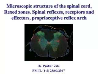
Microscopic structure of the spinal cord, Rexed zones. Spinal reflexes, receptors and effectors, proprioceptive reflex arch Dr. PuskárZita EM II. (1-8) 28/09/2017
Spinal cord in the spinal/vertebral canal intumescentia cervicalis intumescentia lumbalis caudaequina
Spinalganglion/Dorsalrootganglion Dorsal root ganglion (DRG) Spinal nerv Dorsal root Sensory pathway! Sensory neuron: pseudiunipolar cell Ventral root Motor pathway! Sensory neuron Satellite (supporting) cell
Cross section of the spinal cord posterior median sulcus posterior lateral sulcus dorsal horn lateral horn dorsal funiculus dorsal horn white matter gray comissure lateral funiculus gray matter central canal white comissure anterior median fissure ventral funiculus
Neuron types Spinal interneuron (internuncial IN): forms circuits within the spinal cord. Local IN synapses with nearby neurons. ComissuralIN sends axons to the contralateral side of the spinal cord. Propriospinal IN forms synapses with neurons in different spinal segments. Spinal projection neuron (funicular neuron ): its axon travels in the white matter to higher centres Spinal motoneuron (radicular neuron): its cell body is located in the spinal grey matter and its axon projects outside of the spinal cord to innervate effector organs (eg. muscles or vegetative neurons)
Motoneurons human rat Motoneuronsarethe most obviouscelltypes in thespinalgraymatter.
Rexed zones-laminae human rat The gray matter of the spinal cord was divided on the basis of cytoarchitecture of spinal neurons (size, shape, density) into 10 zones or laminae by Rexed. Numbered from dorsal to ventral. Terminology: Lamina I (LI)-Marginal zone, L II-Substantia gelatinosa, L III, IV- no special name, L V- Neck of the dorsal horn, L VI- Base of the dorsal horn, L VII- Intermediate grey, L VIII- no specific name, L IX- Motor nuclei, L X- Central grey
Nuclei of the spinal cord Spinalnuclei: clusters of neurons
Sensation - responses Braincentres Spinalcord somatosensory afferents viscerosensory afferens visceromotor efferents somatomotor efferents Spinal reflex arch receptor effector stimuli skin, muscles tendons viscera glands, smooths muscles striated muscles
Reflex Reflex is an automatic, involuntary and rapid response to a particular stimulus. The basic unit of the reflex is the reflex arch. Whydo we need reflexes? „Sometimesconsciousaction is tooslowtopreventharm”
Components of a reflex arch • Receptor: converts specific stimuli into receptor potential (RP) that reaching its threshold triggers action potentialortransmitterrelease. (exteroceptor: receives stimuli from external environment, proprioceptor: receives stimuli about the position of the body, interoceptor: receives stimuli from internal environment). • Afferent/Sensory neuron : transmit action potential into the CNS • Integration centre: Cranial (brain) or spinal (spinal cord). If the sensory afferent synapses directly on the motoneuron than the reflex is monosynaptic. If the sensory neuron makes synaptic contact with an interneuron that activates other interneurons or motoneurons than the reflex is polysynaptic. • Efferent: motoneuron that innervates the effector • Effector (muscle or gland) „contractsorrelax” in response to the efferent stimuli.
Receptors Primary sensory epithelial cells Secondary sensory epithelial cells Specialized nerve endings
Stretch receptors: muscle spindle and tendon organ extrafusal muscle fibre (innervated by α-motoneuron) Intrafusal muscle fibre (innervated by γ-motoneuron) Muscle spindle: modified intrafusal muscle fibres wrapped around by sensory nerve endings and innervated by γ-motoneurons. Connective tissue capsule separates them from the extrefusal muscle fibers. Golgi tendon organ: encapsulated heavily branched sensory nerve ending running among collagen fibres in tendons. It protects from the overstretching.
Muscle spindle (proprioceptor) It is located in deep within the striated muscle parallel to the extrafusal muscle fibers. It consists of two types of intrafusal muscle fibers : (1-2) nuclear bag fibres (having clusters of nuclei in equatorial region) and (5-7) nuclear chain fibres(nuclei are spread out resembling a chain) Annulospiralending: Ia afferents that wrap around the chain and bag fibres in the equatorial region (ER). Stimuli: change in muscle length and rapid movement/stretching ER. Secondary afferents: Flower spray/Group II afferenswrap around the chain and the static bag fibres in the juxta-equatorial region (JER). Stimuli: constant muscle length. γ motor axon – innervates the polar region (PR) of the intrafusalfibres. (IfthePRscontract → ER is stretched.) It controls the sensitivity of the spindle. ER JER PR
Histology of intrafusal muscle fibres bag chain
Classification of primary afferents bag chain
Proprioceptive reflex archSynonyms: myotatic, monosynaptic, stretch reflex Example: patellar or knee jerk reflex extensors flexors Ia Ia inhibitory Reciprocal inhibition: Ia inhibitory interneurons receiving input from the collaterals of Ia afferents inhibit antagonist motoneurons. Stretching a muscle results in its contraction providing automatic regulation of skeletal muscle length and the posture of the body.
Proprioceptive reflex arch • A proprioceptive reflexes protect the muscles from the passive forces (elongation and stretch), helps the body maintain its balance. • Patella reflex: A sharp tap on the patellar tendon results in sudden kicking movement of the leg. • Receptor: Change in muscle length and rate is the adequate stimulation of the muscle spindle. (Intrafusal fibres in the spindle are lengthened and sensory nerve endings are activated). • Afferents: Ia or II afferents convey information about the change in length and rate of change in length of the muscles. • Integration centre: Spinal cord. Ia/II afferents make monosynaptic connections with motoneurons activating muscles (eg. quadriceps) and their collaterals terminate on Ia inhibitory interneurons located in neighbouring segments and inhibit the activity of motoneurons of antagonist muscles (reciproc inhibition). • Efferents: axons of α-motoneurons • Effectors: muscles (contract and in default of innervation antagonist muscles relax)
Object held in hands stretches the flexors of the arm and due to the proprioceptive reflex arc the flexors will contract and hold the weight. Knee joints tend to flex under the weight of the body, but stretching of the quadriceps femoris muscle activates the proprioceptive reflex which increases the tone of the extensors avoiding the collapse of the human body. Proprioceptive reflex is characteristic for antigravity muscles (flexors of the upper and extensors of the lower limb)! by Dr Kozsurek
Fine tuning • γ-loop • Renshaw cell • Golgi tendon reflex
γ-motoneuron, γ-loop and α-γ co-activation • The sensitivity of the muscle spindle is adjusted through gamma efferent innervation. γ-motorneurons contract intrafusal muscle fibres and make the spindle more sensitive. • A muscle contraction can be evoked in two different ways: • Directly, through α-motoneurons. • Indirectly, through the activation of the γ-loop and the proprioceptive reflex arc. • γ-loop: descending pathways can stimulate gamma-motoneurons which innervate intrafusal muscle fibres. Contracting intrafusal fibres shorten the muscle spindle itself which has a similar effect to that when the extrafusal fibres around the spindle are passively stretched. This stretch(-like) signal evokes the spinal proprioceptive reflex activating α-motoneurons, and finally contracting extrafusal fibres. • α -γ coactivation: corticospinal fibers activate both α- and γ-motoneurons that allows Iafibers to continue to sense muscle length during contraction.
Recurrent inhibition by Renshaw cells Renshaw cell is an inhibitory interneuron that receives direct input from α-motoneurons via axon collaterals and inhibits motoneurons in synergistic motor pool (recurrentinhibition), Ia inhibitory interneurons and other Renshaw cells. Depending on the situation, γ-motoneurons can also be inhibited by Rensaw cells. The major function of the Renshaw cell is modulation of motor output and protection from over-excitation.
Golgi tendon organSlow adapting mechanoreceptor Golgi tendon reflex is a polysynaptic (disynaptic) reflex that protects the muscle from tensing excessively. Receptor: Golgi tendon organ (adequate stimuli: tension during muscle contraction) Afferents: Ib thick myelinated axons Integration centre: Spinal cord (Ib sensory afferents terminate on Ibinhibitoryinterneurons that inhibitα-motoneurons) Efferent:α-motoneuron axon Effector:extrafusalfibres of skeletal muscle (relax)
Muscle spindle and Alzheimer disease? Cheret Cet al: Bace1 and Neuregulin-1 cooperate to control formation and maintenance of muscle spindles EMBO J. 2013 Jul 17;32(14):2015-28. doi: 10.1038/emboj.2013.146. Epub 2013 Jun 21. „The enzyme beta-secretase generates amyloid-beta peptides, the constituents of the damaging amyloid plaques in Alzheimer’s disease. In order to halt the disease, researchers are developing drugs aiming to block the enzyme. …Together with the growth factor Neuregulin-1, Bace1 is essential for the formation and maintenance of the function of muscle spindles, which ensure a constant muscle tone and protect muscles from overstretching. ” https://www.mdc-berlin.de