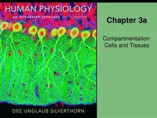
Chapter 3a
Chapter 3a. Compartmentation: Cells and Tissues. About this Chapter. Body compartments Biological membranes Intracellular compartments Tissue types and characteristics Tissue remodeling Organs. Three Major Body Cavities. POSTERIOR. ANTERIOR. Cranial cavity. Pleural sac. Thoracic

Chapter 3a
E N D
Presentation Transcript
Chapter 3a Compartmentation: Cells and Tissues
About this Chapter • Body compartments • Biological membranes • Intracellular compartments • Tissue types and characteristics • Tissue remodeling • Organs
Three Major Body Cavities POSTERIOR ANTERIOR Cranial cavity Pleural sac Thoracic cavity Pericardial sac Diaphragm Abdominal cavity Abdominopelvic cavity Pelvic cavity Figure 3-1
Lumens of Hollow Organs • Hollow organs • Heart • Lungs • Blood vessels • Intestines • Lumen • Not the internal environment
Functional Compartments • Outside Body • Extracellular fluid • Plasma • Interstitial fluid • Intracellular fluid • Organelles and vacuoles
Body Fluid Compartments Cell membrane Capillary wall Blood cells Blood vessel Plasma Interstitial fluid Intracellular fluid ECF ICF Cell membrane Figure 3-2
Cell Membrane: Overview • Membranes in the body Loose connective tissue Pericardial membrane Cell Heart The pericardial membrane is a tissue that surrounds the heart. Seen magnified, the pericardial membrane is a layer of flattened epithelial cells supported by connective tissue. Each cell of the pericardial membrane has a cell membrane surrounding it. The cell membrane is a phospholipid bilayer. Figure 3-3
Cell Membrane: Functions • Physical barrier • Gateway for exchange • Communication • Cell structure
Cell Membrane: Structure • The fluid mosaic model of a biological membrane Carbohydrate group of glycoprotein Carbohydrate group of glycolipid Extracellular surface of membrane Membrane splits into layers in freeze-fracture electron microscopy. Proteins Intracellular surface of membrane Lipid tails form the interior layer of the membrane. Cholesterol molecules insert themselves into the lipid layer. Phospholipid heads face the aqueous intracellular and extracellular compartments. Figure 3-4
Proteins Integral Peripheral Lipid-anchored Cell Membrane: Composition • Lipids • Phospholipids • Sphingolipids • Cholesterol
Cell Membrane: Composition Table 3-1
Cell Membrane: Structure and Formation • Phospholipids have polar and non-polar regions Phospholipid molecules have polar heads and nonpolar tails. The “R” group is a variable polar group. (a) Polar head (hydrophilic) Nonpolar fatty acid tail (hydrophobic) Structural model Molecular models Stylized model Figure 3-5a
Cell Membrane: Formation • Membrane phospholipids form bilayers, micelles, or liposomes Phospholipids arrange themselves so that their nonpolar tails are not in contact with aqueous solutions such as extracellular fluid. (b) Tails Phospholipidbilayer forms a sheet. Micelles are droplets of phospholipids. Liposomes have an aqueous center. Figure 3-5b
Cell Membrane: Proteins • The three types of membrane proteins: integral, peripheral, and lipid-anchored Glycoprotein Integral (transmembrane) protein Peripheral protein Lipid-anchored proteins Peripheral protein Cytoskeleton proteins Cytoplasm Figure 3-6
Cell Membrane: Lipid Rafts • Sphingolipids and alkaline phosphatase Figure 3-8
Cell Membrane Components CELL MEMBRANE consists of Cholesterol Phospholipids, Sphingolipids Carbohydrates Proteins together form together form together form Glycolipids Glycoproteins Lipid bilayer functions as whose functions include Selective barrier between cytosol and external environment Structural stability Cell recognition Immune response Figure 3-9
Intracellular Compartments • Cytoplasm • Cytosol • Inclusions • Organelles • Nucleus
Cell Compartments THE CELL • A map for the study of cell structure is composed of Cell membrane Nucleus Cytoplasm Membranous organelles Cytosol Inclusions • Mitochondria • Endoplasmic reticulum • Golgi complex • Lysosomes • Peroxisomes • Lipid droplets • Glycogen granules • Ribosomes • Vaults • Proteasomes • Cytoskeleton • Centrioles • Centrosomes • Cilia • Flagella Extracellular fluid Figure 3-11
Inclusions Have No Membranes • Ribosomes • Free • Fixed • Polyribosomes • Proteasomes • Vaults • RNA/protein
Cytoplasmic Proteins Fibers • Actin (microfilaments) • Intermediate • Myosin • Keratin • Neurofilaments • Microtubules • Tubulin • Centrioles, cilia, flagella
Microtubule function • Centrioles • Pull chromosomes • Form core in cilia • Cilia and flagella • Fluid movement
Centrioles Figure 3-13a–b
Cilia and Flagella Figure 3-13c–d
Cytoskeleton: Function • Cell shape • Internal organization • Intracellular transport • Assembly of cells into tissues • Movement
Cytoskeleton and Cytoplasmic Protein Fibers Microvilli increase cell surface area. They are supported by microfilaments. Microfilaments form a network just inside the cell membrane. Microtubules are the largest cytoskeleton fiber. Intermediate filaments include myosin and keratin. (a) (b) Figure 3-14
Cytoskeleton and Cytoplasmic Protein Fibers • Motor proteins move on cytoskeletal fibers Organelle Motor protein ATP Direction of movement Cytoskeletal fiber Figure 3-15
Mitochondria • Membrane-enclosed compartments • Unique DNA • Site of cellular ATP generation
Mitochondria Cytoplasm of cell Inner membrane Matrix Matrix is the innermost compartment. Outer membrane Cristae The intermembrane space forms a compartment. Cytosolic side of membrane Figure 3-16
Endoplasmic Reticulum (ER) • Smooth ER • Synthesis of fatty acids, steroids, lipids • Modified forms in liver, kidney, muscles • Rough ER • Rows of ribosomes • Protein assembly and modification
Endoplasmic Reticulum Lumen of endoplasmic reticulum Ribosomes are attached to cytosolic side of rough endoplasmic reticulum. Smooth endoplasmic reticulum Endoplasmic reticulum Figure 3-17