Download
1 / 1
20 likes | 235 Vues
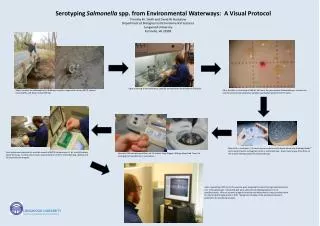
Serotyping Salmonella spp. from Environmental Waterways: A Visual Protocol Timothy M. Smith and David W. Buckalew Department of Biological and Environmental Sciences Longwood University Farmville, VA 23909. An example of (+) agglutination from this experiment.

E N D
SerotypingSalmonella spp. from Environmental Waterways: A Visual Protocol Timothy M. Smith and David W. Buckalew Department of Biological and Environmental Sciences Longwood University Farmville, VA 23909 An example of (+) agglutination from this experiment Courtesy of Oxoid™ website Upon returning to the laboratory, samples are processed via membrane filtration. Water Samples are collected from 3 different locations: Appomattox River (APP2), Sayler’s Creek (SAY5), and Green Creek (GRE16). After the filter is incubated in BGB for 24 hours, the presumptive Salmonella spp. colonies are enumerated and representative samples aseptically transferred to TSI slants. + After 48 hrs. incubation, TSI results are recorded and the Rapid Salmonella Antibody Beads™ test is performed to serologically confirm Salmonella spp. Gram-staining was then done on the isolates testing positive for Salmonella spp. Each isolate was prepared for multiple rounds of MPCR to determine O, H1, and H2 antigen allelic formulae. Control primers were used to further confirm Salmonella spp. identity and for experimental integrity. Genomic DNA was extracted from all 32 isolates using Qiagen’sDNeasy Blood and Tissue kit according to manufacturer’s instructions. Upon completing a PCR cycle, the samples were prepared for pulsed-field gel electrophoresis on a 1.5% agarose gel. Completed gels were visualized and photographed on a UV transilluminator. After an isolate’s antigenic formulae was determined, it was correlated with the World Health Organization’s 2007 “Antigenic Formulae of the Salmonella Serovars” publication for serotyping analysis. LONGWOOD UNIVERSITY Department of Biological and Environmental Sciences