Anorectal diseases
430 likes | 670 Vues
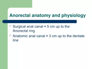
Anorectal diseases. Ahmad kachooei Assistant Professor of Qom Medical University. Anatomy. Any patient with anal/ perianal symptoms requires a careful history and physical, including a digital rectal examination

Anorectal diseases
E N D
Presentation Transcript
Anorectal diseases Ahmad kachooei Assistant Professor of Qom Medical University
Any patient with anal/perianal symptoms requires a careful history and physical, including a digital rectal examination • defecography, manometry, CT scan, MRI, contrast enema, endoscopy, endoanal ultrasound, or examination under anesthesia may be required to arrive at an accurate diagnosis
Hemorrhoids • Hemorrhoids are cushions of submucosal tissue containing venules, arterioles, and smooth-muscle fibers . • Three hemorrhoidal cushions : • left lateral • right anterior • right posterior
Hemorrhoidal Function • continence mechanism • aid in complete closure of the anal canal at rest • Because hemorrhoids are a normal part of anorectal anatomy, treatment is only indicated if they become symptomatic.
Etiology • Excessive straining • increased abdominal pressure • hard stools
Types • External hemorrhoids are located distal to the dentate line and are covered with anoderm • Internal hemorrhoids are located proximal to the dentate line and covered by insensate anorectal mucosa
Internal hemorrhoids • Graded according to the extent of prolapse: • First-degree hemorrhoids bulge into the anal canal and may prolapse beyond the dentate line on straining. • Second-degree hemorrhoidsprolapse through the anus but reduce spontaneously
Third-degree hemorrhoids prolapse through the anal canal and require manual reduction. • Fourth-degree hemorrhoidsprolapse but cannot be reduced and are at risk for strangulation
Combined internal and external hemorrhoids • Combined internal and external hemorrhoids straddle the dentate line and have characteristics of both internal and external hemorrhoids • Hemorrhoidectomy often is required for large, symptomatic, combined hemorrhoids.
Postpartum hemorrhoids • result from straining during labor, which results in edema, thrombosis, and/or strangulation. • Hemorrhoidectomy is often the treatment of choice, especially if the patient has had chronic hemorrhoidal symptoms
Treatment • Medical Therapy: • Bleeding from first- and second-degree hemorrhoids often improves with the addition of dietary fiber, stool softeners, increased fluid intake, and avoidance of straining • Associated pruritus may often improve with improved hygiene
Treatment • Rubber Band Ligation • Infrared Photocoagulation • Sclerotherapy • Excision of Thrombosed External Hemorrhoids
Operative Hemorrhoidectomy • Closed SubmucosalHemorrhoidectomy • Open Hemorrhoidectomy • Whitehead's Hemorrhoidectomy • Procedure for Prolapse and Hemorrhoids/Stapled Hemorrhoidectomy(PPH)
Complications of Hemorrhoidectomy • Postoperative pain • Urinary retention • fecal impaction • Bleeding • Infection • Incontinence • anal stenosis • ectropion (Whitehead's deformity)
Anal Fissure • A fissure in ano is a tear in the anoderm distal to the dentate line. • The vast majority of anal fissures occur in the posterior midline. Ten to 15% occur in the anterior midline. • Less than 1% of fissures occur off midline
pathophysiology of anal fissure • trauma from either the passage of hard stool or prolonged diarrhea. • This cycle of pain, spasm, and ischemia contributes to development of a poorly healing wound that becomes a chronic fissure.
Symptoms and Findings • extremely common • tearing pain with defecation and hematochezia (usually described as blood on the toilet paper) • painful anal spasm lasting for several hours after a bowel movement
physical examination • can be seen in the anoderm by gently separating the buttocks • Patients are often too tender to tolerate digital rectal examination
Types • An acute fissure is a superficial tear of the distal anoderm and almost always heals with medical management • Chronic fissures develop ulceration and heaped-up edges with the white fibers of the internal anal sphincter visible at the base of the ulcer. • There often is an associated external skin tag and/or a hypertrophied anal papilla internally.
A lateral location of a chronic anal fissure may be evidence of an underlying disease such as Crohn's disease, HIV, syphilis, tuberculosis, or leukemia. • If the diagnosis is in doubt or there is suspicion of another cause for the perianal pain, such as abscess or fistula, an examination under anesthesia may be necessary
Treatment • Therapy focuses on breaking the cycle of pain, spasm, and ischemia • First-line therapy to minimize anal trauma includes bulk agents, stool softeners, and warm sitz baths • The addition of 2% lidocaine jelly or other analgesic creams can provide additional symptomatic relief.
Treatment • Nitroglycerin ointment (0.2%) • Both oral and topical calcium channel blockers (diltiazem and nifedipine) • arginine (a nitric oxide donor) • topical bethanechol (a muscarinic agonist) • Medical therapy is effective in most acute fissures, but will heal only approximately 50 to 60% of chronic fissures.
Botulinum toxin (Botox) Surgical therapy: traditionally has been recommended for chronic fissures that have failed medical therapy • Approximately 30% of the internal sphincter fibers are divided laterally • Healing is achieved in more than 95% of patients by using this technique
Anorectal Sepsis and Cryptoglandular Abscess • The majority of anorectalsuppurative disease results from infections of the anal glands (cryptoglandular infection) • Their ducts traverse the internal sphincter and empty into the anal crypts at the level of the dentate line
perianal and perirectal spaces • perianal space • intersphincteric space • ischiorectal space (ischiorectalfossa) • supralevator spaces
Abscess • perianal abscess • ischiorectal abscess • Intersphincteric abscesses • supralevator abscesses
Diagnosis • Severe anal pain • Walking, coughing, or straining can aggravate the pain • Palpable mass • fever • urinary retention • life-threatening sepsis
Treatment • Drainage as soon as the diagnosis is established • diagnosis is in question:an examination under anesthesia • Antibiotics : • extensive overlying cellulitis • Immunocompromised • diabetes mellitus • valvular heart disease
Fistula in Ano • Drainage of an anorectal abscess results in cure for about 50% of patients. • The fistula usually originates in the infected crypt (internal opening) and tracks to the external opening, usually the site of prior drainage.
Diagnosis • Patients present with persistent drainage from the internal and/or external openings. • An indurated tract often is palpable • Goodsall's rule can be used as a guide in determining the location of the internal opening .
intersphincteric fistula tracks through the distal internal sphincter and intersphincteric space to an external opening near the anal verge
transsphincteric fistula often results from an ischiorectal abscess and extends through both the internal and external sphincters
suprasphincteric fistula originates in the intersphincteric plane and tracks up and around the entire external sphincter
extrasphincteric fistula originates in the rectal wall and tracks around both sphincters to exit laterally, usually in the ischiorectalfossa
Treatment • The goal of treatment of fistula in ano is eradication of sepsis without sacrificing continence. • Simple intersphincteric fistulas often can be treated by fistulotomy (opening the fistulous tract), curettage, and healing by secondary intention
Treatment • seton : • Cutting setons • noncuttingseton • endorectal advancement flap :Higher fistulas • Fibrin glue