Download
1 / 16
210 likes | 586 Vues
Hyaluronic acid-based scaffolds for repair strategies after spinal cord injury: A behavioral study. Zin Z. Khaing 1 , Sydney A. Geissler 1 , Sandra V. Aguilar 1 , Timothy Schallert 2 and Christine E. Schmidt 1.

E N D
Hyaluronic acid-based scaffolds for repair strategies after spinal cord injury: A behavioral study Zin Z. Khaing1, Sydney A. Geissler1, Sandra V. Aguilar1, Timothy Schallert2 and Christine E. Schmidt1 1The University of Texas at Austin, Department of Biomedical Engineering2The University of Texas at Austin, Department of Psychology
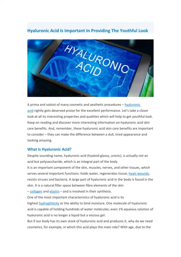
Hyaluronic Acid (HA) • Extracellular and cell surface-associated • Highly viscous and hydrophilic • Can modulate cell behavior • (development, inflammation axonal growth) • Vital role in scar-free wound healing
Hyaluronic Acid Materials Vitreous substitute (Healon®) Viscosupplementation (Synvisc®) Other Applications Dermal Filler (Restylane®), Adhesion Barrier (Seprafilm®) In Our Group Images from healon.com and synvisc.com
After Acute Spinal Cord Injury Acute Phase • Cell death • Axonal damage • Local hemorrhage • Inflammation and edema Sub-acute Phase • Expansion of injured area during secondary phase • Activation of microglia and macrophages • Astrocyte activation • Scar formation (involve fibroblasts, meningial cells, infiltrating progenitors and astrocytes) production of glial scar including CSPGs
HA after SCI Previous studies from our lab showed that the presence of high MW HA hydrogel can • Limit macrophage and microglia infiltration after SCI • Limit astrocyte response in vivo and astrocyte proliferation in vitro • Can alter the amount of CSPG deposition • Support axonal outgrowth after SCI
HA hydrogels after SCI Spinal Cord Injury Model: Cervical lateral hemisection at C3/C4 The most clinically relevant model (63% of human SCI are at the cervical level) Rodents mainly use their forelimbs, therefore, behavioral deficits and improvements can be assessed more readily in an accurate manner Hydrogels Used: GMHA, LN/GMHA, LN/gelfoam, gelfoam, and laminectomy (n=6 in each group) Analysis: (a) Behavioral (b) Histological
Behavioral Tests • Forelimb Locomotor Score (FLS) • General forelimb usage during locomotion will be scored. • Non-linear scale from 0-17. • 2. Cylinder • Forelimb exploration will be assessed. • The numbers of forelimb contacts (left, right, and both) with the cylinder walls were counted and expressed as a percentage of total placements. • 3. Forelimb placing • Forelimb placing tests response to sensorimotor/proprioceptive detection of edge of table with capacity vibrissae (whiskers) • 4. Adhesive removal (Sensory bias) • Small adhesive backed (sticky) labels are placed on the distal–radial aspect of both forelimbs • Contact and removal times are recorded • Tests preference for responding sensory stimuli as well as motor coordination
Behavioral Tests • Forelimb Locomotor Score (FLS) • General forelimb usage during locomotion will be scored. • Non-linear scale from 0-17. • 2. Cylinder • Forelimb exploration will be assessed. • The numbers of forelimb contacts (left, right, and both) with the cylinder walls were counted and expressed as a percentage of total placements. • 3. Forelimb placing • Forelimb placing tests response to sensorimotor/proprioceptive detection of edge of table with capacity vibrissae (whiskers) • 4. Adhesive removal (Sensory bias) • Small adhesive backed (sticky) labels are placed on the distal–radial aspect of both forelimbs • Contact and removal times are recorded • Tests preference for responding sensory stimuli as well as motor coordination
Cylinder Height • LN-soaked gelfoam implants showed the most improved behavior
Paw Preference (Cylinder) • Implanted animals showed some increase in affected limb usage at 12 week post surgery.
Behavioral Tests • Forelimb Locomotor Score (FLS) • General forelimb usage during locomotion will be scored. • Non-linear scale from 0-17. • 2. Cylinder • Forelimb exploration will be assessed. • The numbers of forelimb contacts (left, right, and both) with the cylinder walls were counted and expressed as a percentage of total placements. • 3. Forelimb placing • Forelimb placing tests response to sensorimotor/proprioceptive detection of edge of table with capacity vibrissae (whiskers) • 4. Adhesive removal (Sensory bias) • Small adhesive backed (sticky) labels are placed on the distal–radial aspect of both forelimbs • Contact and removal times are recorded • Tests preference for responding sensory stimuli as well as motor coordination
Acknowledgements • Dr. Christine Schmidt • Sydney Geissler • Entire Schmidt lab Collaborators • Dr. Timothy Schallert, UT-Austin • Raymond Grill, UT-Houston Supported By Texas Paralysis Foundation David Van Wagner Foundation Gillson Longenbough Foundation