Download
1 / 77
790 likes | 2.03k Vues
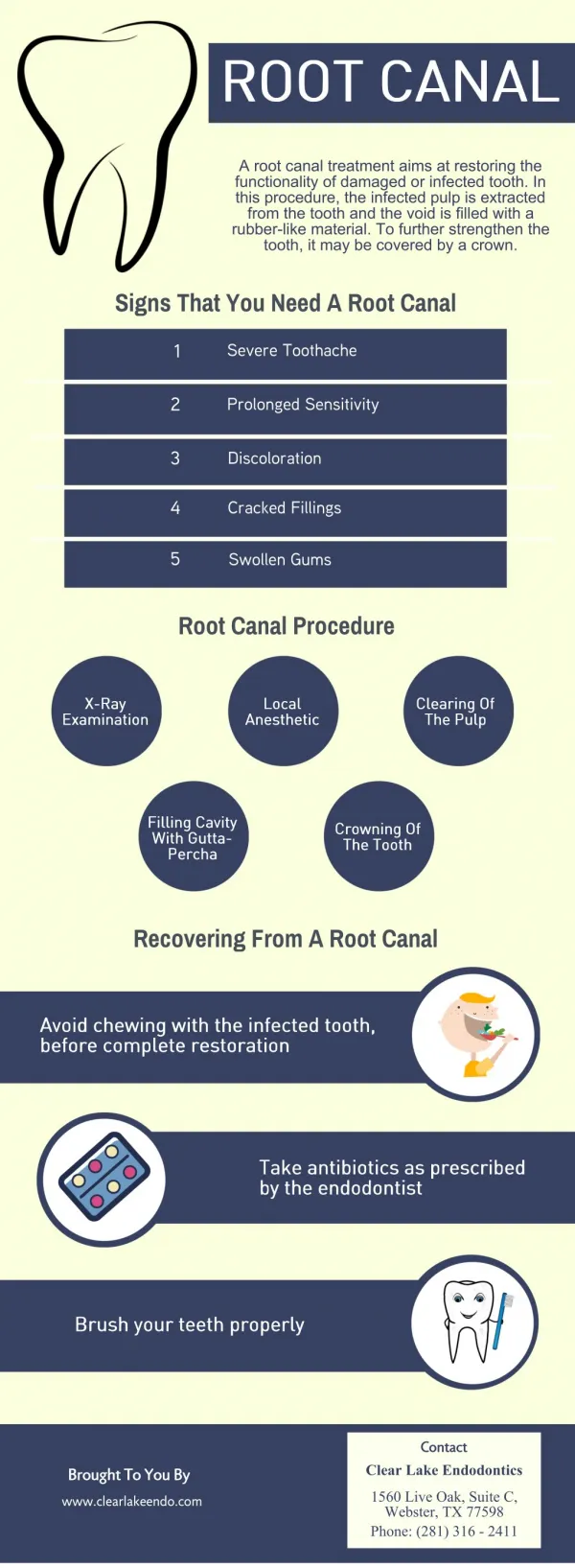
Learn about the root canal procedure, causes of pulp damage, and signs of damaged pulp. Find out how the GentleWave system cleans and disinfects root canals effectively.

E N D
PURPOSE OF THE PRESENTATION:1. Inform you about the root canal procedure.2. Help answer any questions.3. Make the x-rays more understandable.
RUNNING THE PRESENTATION:1. The assistant will start the slide show for you.2. Press the DOWN ARROW key to move to the next slide.3. Press the UP ARROW key to move back
RUNNING THE PRESENTATION:1. When it finishes the last slide says “THE END”2. Please let the staff know when you are done.
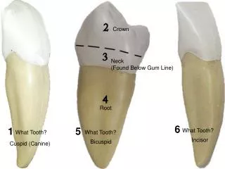
Crown A healthy tooth consistsof a crown...
Crown Root ...and a root.
Under the hard layers of enamel and dentin... Enamel Dentin
Pulp Tissue is soft tissue called pulp. Enamel Dentin
PULP CONTAINS: • Specialized tooth cells • These can calcify the tooth. • Connective tissues • Arteries / Veins • Nerve tissue • The pulp is often called “The NERVE” of the tooth.
The pulp creates dentin during tooth development.(Ages 2-18)
Damaged Pulp The pulp maybecome infected or damaged. Decay
The Pulp (“Nerve”) can become damaged or infected from: • Injury or trauma to the tooth. • UNTREATED deep decay into or near the pulp. • Deep decay which required deep FILLINGS or a crown to protect the tooth.
Damaged Pulp Abscess If left untreated,an abscess will form. Decay
How do “deep fillings” cause pulp problems?? • Deep decay leads to deep fillings. • A crown is sometimes needed to protect the tooth from splitting. • These repairs will sometimes lead to pulp “scarring.”
How do “deep fillings” cause pulp problems?? • “Scarring” of the pulp may show on the x-ray as “calcifications” or hard tooth deposits inside the pulp chamber. • This limits blood supply and the pulp dies.
Signs of damaged pulp: • May start with a hot or, more commonly, cold sensitivity. • Sometimes a sharp pain, throbbing pain, or spontaneous pain for no reason at all.
Signs of damaged pulp: • As the inflammation moves outside your tooth, it will become tender to biting pressure, or touch. • Next a tenderness develops around the tooth and deep in the jaw.
Signs of damaged pulp: • Bone “eaten away” (a dark spot on the end of the root) is the first sign seen on an X-Ray. • Your symptoms are important in making the earliest diagnosis possible.
Signs of damaged pulp: • Next an abscess may form! This is often seen as a swelling and tenderness in the gums. • When bone is eaten away around the end of your tooth, portions of the tooth may also be destroyed.
Sometimes there areno symptoms. Some endodontic problems are only evident on an x-ray... This is just one of the reasons why it is SO important for us to take x-rays.
During your TREATMENT visit at our office, the following steps are taken:
Preparation for Treatment • The tooth is examined. • Additional X-rays may be indicated. • Anesthetic is given. • A dental dam is placed.
A dental dam is a small sheetof rubber that surroundsthe tooth. It will isolate your tooth and keep it clean during the procedure. As well as keep the debris out of your mouth during treatment.
…I will make an openingin the tooth or crown... Access Opening
A microscope may be used to see more clearly deep inside the tooth. This allows for a much clearer view when there are potential problems.
…then I gently clean the pulp from the pulp chamber... Access Opening Cleaned and Shaped Pulp Chamber
...and canals inside the roots. Access Opening Cleaned and Shaped Pulp Chamber and Root Canal
Before I clean the canals I determine the length of your tooth using very small, thin instruments called FILES. The files look like a small thin wire… it simply has file edges along the outside.
The first ones used are 10/100th of a millimeter.That is 1/254th of an inch!! So small they may occasionally separate or break within the tooth. This rarely causes a problem.
IF a file were to break, they are so fine, they are often sealed within the canal. It is not a cause for concern. I will certainly advise you, if any difficulties occur during the procedure.
After I measure your tooth I will begin to clean it out with The GentleWave system.
The GentleWave uses multisonic cleaning technology and various cleaning fluids to remove debris from inside your tooth.
While it cleans with these solutions it also simultaneously vacuums them back out along with the debris leaving your tooth VERY clean.
It even cleans microscopic areas that we would be unable to reach using conventional cleaning methods.
The cleaning fluids travel in and out of your tooth through a special designed hand piece with a nozzle on the end that rests in your tooth.
A temporary BLUE seal is placed on your tooth to hold the hand piece in place and keep the nozzle centered. SEAL NOZZLE
The nozzle is placed inside your tooth. The GentleWave will then ultrasonically clean your tooth out.
Once the root canals are cleaned, shaped, and disinfected...
…I willfill the canals... Filled Root Canals
…with a rubber-like material called gutta-percha. Filled Root Canals
At this time the root canal is FINISHED, and the tooth must be sealed Three kinds of sealing techniques may be used.
The ways of sealing yourROOT CANAL are: • A Temporary seal. • A Base (core) for a new crown. • The Repair of an existing crown.
Temporary SEAL A temporary seal isNOTa filling. Filled Root Canals
Temporary Seal This seal is designed to ONLY last 2 to 4 weeks. Filled Root Canals
Temporary Seal With a Temporary Seal you are NOT finished with your care! Filled Root Canals
They will remove the seal, place a final restoration, and begin your treatment for a CROWN. Within 4 weeksof your root canal, you MUSTreturn to your general dentist.
CROWN REPAIR If your tooth already has a crown, this will seal the opening.