Download
1 / 24
250 likes | 372 Vues
This comprehensive guide explores the anatomy of the respiratory system across several animal species, including carnivores, bovines, equines, avians, and porcine. It details the structure and functions of the nares, nasal cavity, paranasal sinuses, pharynx, larynx, and lower respiratory system, emphasizing their roles in heating, moistening air, and protecting against particles. Additionally, it discusses species-specific adaptations and the importance of the vomeronasal organ and guttural pouch in particular species, enhancing our understanding of veterinary medicine.

E N D
Respiratory System The University of Nottingham School of Veterinary Medicine and Science Created by Chloe Hughes Veterinary Student
Nares (nostrils) The nares are surrounded by hairless skin and supported by nasal cartilages Species differences: • Carnivores: a nasal plate that is divided by a medium groove called the philtrum. • Bovine: a nasolabial plate that has cornified stratified epithelium and serous glands • Equine: have an incomplete cartilaginous ring allowing distensible nostrils • Avian: slit-like openings with an overhanging operculum. • Porcine: snout contains os rostrale (bone)
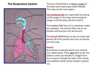
Upper Respiratory System Consists of the nasal cavity, paranasal sinuses, nasopharynxand larynx. Functions: heating/moistening air and defence against incoming particles. Nasal Cavity Nasopharynx Hard Palate Oesophagus Oropharynx Larynx Oral Cavity Trachea Tongue Laryngopharynx Epiglottis Soft Palate
Nasal Cavity • Paired chambers separated by a nasal septum. • Connected anteriorly to the nares and posteriorly to the nasopharynxthrough choanae. • Divided into meatus by nasal conchae that consist of turbinate bone and the covering respiratory mucosa (pseudostratified columnar epithelium and the underlying lamina propria) • Allows a defence (sneeze reflex and cilia) and warming and moistening function Dorsal conchae Caudal conchae Vestibule Choanae Ventral conchae
Nasal Cavity Dorsal meatus The nasal conchae divide the nasal cavity into meatus. In the horse: • Dorsal meatus leads to the olfactory mucosa • Middle meatus leads to the paranasal sinuses • Ventral and the common meatus lead to the pharynx The nasal cavity anatomy is fairly simple in horse compared to the complex structure of the canine nasal cavity. Middle meatus Ventral meatus Common meatus Nasal Septum
Vomeronasal Organ • In addition to the olfactory cells at the end of the dorsal meatus, there is also an accessory olfactory structure, the vomeronasal organ. • The vomeronasal organ consists of paired blind ending ducts contained within the hard palate. It connects the nasal and oral cavities and contains unique chemoreceptors. • Functions: • Pheromone detection • Flehmen response Vomeronasal Organ
Paranasal Sinuses Air filled sinuses connected to the nasal cavity through the middle meatus. Functions include resonating (voice), insulation and cooling of the brain and light weight skull construction. The frontal and maxillary sinuses are common to all species. The frontal sinus drains into the ethmoidal meatus (except in the horse).It has a rostral and caudal part, with the caudal part communicating with horn in cattle. Maxillary sinus is divided into two compartments and drains into the middle meatus.It contains the cheek teeth (molars and premolars). Cattle have palatine, sphenoid, lacrimal and palatomaxillary sinuses Birds have an infraorbital sinus below the eye Frontal sinus Sphenopalatine sinus Rostral maxillary sinus Caudal maxillary sinus
Pharynx The pharynx connects the oral cavity and nasal cavity to the trachea and oesophagus. It functions as a passageway for air and food but also as a resonating chamber. It is split into three regions, the nasopharynx, oropharynx and laryngopharynx. Nasopharynx Laryngopharynx Oropharynx
Eustachian tube and Guttural Pouch The Eustachian tubeconnects the nasopharynx to the middle ear. In the horse, an out pouching forms the guttural pouch. The guttural pouch is approximately a 300-500cm³ space in the equine skull, between the skull base, atlas, pharynx and oesophages.It is divided into medial and lateral compartments by the stylohyoid bone. It functions in cooling of the blood entering the brain.
Larynx The larynx is a structure connecting the pharynx to the trachea and oesophagus. It is suspended by the hyoid bones that articulate with the base of the skull. Functions: • Phonation • Swallowing • Breathing It consists of hyaline and elastic cartilages: cricoid, thyroid, epiglottis and 2 arytenoid cartilages. The larynx contains the vocal cords required for phonation. In birds, the larynx consists of only cricoid and arytenoid cartilages. It does not have vocal folds therefore sound is created by the syrinx at the tracheal bifurcation
Larynx and Hyoid Apparatus Equine larynx and hyoid apparatus Arytenoid cartilage Epiglottis Stylohyoid Epihyoid (vestigial) Cricoid cartilage Ceratohyoid Thyrohyoid Basohyoid Thyroid cartilage
Epiglottis The epiglottis channels food towards the epiglottis during swallowing, preventing ingesta entering the lungs through the trachea The larynx, and most of the epiglottis, is lined with stratified squamous epithelium up to the vocal cords . The rest of the larynx is lined by pseudostratified columnar epithelium, like the respiratory tract.
Lower Respiratory System The lower respiratory system consists of the trachea down to the alveoli. Functions: • Gas exchange • Metabolic functions (surfactant synthesis, histamine synthesis, activation of angiotensin) • Body temperature regulation • Acid-base regulation • Vocalisation Consists of two types of airways: • Conducting – carry, warm, filter and saturate air going to respiratory airways • Respiratory – undergo gaseous exchange
Lungs The lungs obtain their appearance through the deep fissures. Canine: • the right lung is larger and is divided into cranial, middle, caudal and accessory lobes. • The left lung is divided into a cranial and caudal lobe. Equine: • The lungs are more equal in size. • There is no external lobationother than the accessory lobe of the right lung. Bovine: • The right lung is significantly larger than the left • The left lung is divided into a cranial and caudal lobe, with the cranial further divided into two parts. • The right lung is divided into cranial, middle, caudal and accessory lobes
Lower Respiratory System Trachea Bronchus Bronchiole Conducting System Respiratory Bronchiole Alveolar duct Alveolar sac Alveolus Respiratory System Fallow deer lung cast NB. The upper respiratory system is also part of the conducting system
Trachea • Flexible tube held open by cartilaginous c-shaped rings • Rings are joined by connective tissue • Trachealis muscle runs along the dorsal aspect • lined by ciliated columnar epithelium that functions as a ‘mucociliary escalator’ • Mucus traps small particles which are moved towards the pharynx by the cilia, removing debris from the lungs. • Various goblet cells line the trachea, producing the mucous coat. Cartilaginous C- shaped rings
Trachea Ciliated epithelium Glands Cartilage
Conducting System • Bronchi • Lined by pseudostratifiedepitheliumand goblet cells • Developed spiral bands of bronchial smooth muscle regulate the volume of the conducting zone • Outside the smooth muscle are irregular plates of bronchial cartilage • Surrounded by a highly vascular peribronchial sheath that contains lymphatic vessels • Bronchiole • The epithelium is reduced to cuboidal with no mucus secreting glands • Although cartilage is absent, the spiral bronchial muscle is well developed in this region • There is collateral ventilation (connections) between bronchioles, making them less vulnerable to blockage • Lacks the vascular sheath
Conducting System Bundles of smooth muscle Cartilage (hyaline) Bronchus Blood vessel
Respiratory System Respiratory Bronchiole: • Has a few alveolar scattered along its wall • Lined by typical bronchiolar epithelium, but changes to simple squamous at alveolus entrance • The alveoli entrance is guarded by bronchial smooth muscle Alveolar duct: • Have alveoli on its walls with the openings guarded by smooth muscle Alveolar sac: • Usually there are clusters of alveolar sacs on the end of the alveolar duct Alveolus: • Minute polygonal chambers whose diameter changes with breathing • The wall is a thin irregular sandwich with the two outer surfaces formed by epithelial cells and an inner network of capillaries
Respiratory System Alveolar Sac Bronchiole Alveolar Duct
Thoracic cavity and pleura • Intercostal muscles: • External – run in a caudoventral direction and are responsible for inspiration • Internal – run in a caudodorsaldirection and consists of 2 components: the interosseus, responsible for expiration, and interchondral, responsible for inspiration. Pleura: • A serous membrane lining the contents of the thoracic cavity • Consists of a single layer of flat epithelium and underlying propria • The thoracic cavity is lined by parietal pleura. • Each lung is covered in visceral pleura, arranged as closed pleural sacs. • The space between the right and left pleural sacs is the mediastinum • The narrow space between the parietal and visceral pleura is the pleural cavity. This is under sub-atmospheric pressure.
Diaphragm Muscular part Tendinous part • A dome shaped structure separating the thoracic and abdominal cavities. • Convex on its cranial surface. • Has a muscular peripheral part and a tendinous central area. • Muscular part has sections arising from the xiphoid process of sternum, vertebral column and caudal ribs. • Supplied by the phrenic nerve • During inspiration, the diaphragm contracts to increase the volume of the thoracic cavity, thus decreasing its pressure to draw air in. • Vice versa occurs for expiration with the diaphragm relaxing.
Avian Respiratory Tract The avian tract differs in the following ways: • A choana (opening) connecting the nasal and oral cavity • The larynx does not have an epiglottis, so muscles constrict, closing the glottis, to allow passage of food • The trachea is longer and wider than in mammals, and consists of interlocking rigid cartilages. • A syrinx is located at the trachea bifurcation and is responsible for sound production • Air sacs, acting as bellows, may store air during respiration, allowing the lungs to be constantly supplied by fresh air. • The lungs are stiffer and do not expand like mammalian lungs due to more cartilage being present • There is a counter current exchange in the lungs of the bird, allowing for efficient exchange • Birds experience a continuous unidirectional air flow through the lungs Parabronchi