Bleeding in Early Pregnancy
571 likes | 1.82k Vues
Bleeding in Early Pregnancy. Dr/ Sahar Elkheshen Lecturer of maternity and neonatal nursing Faculty of Applied medical sciences MUST. Objectives : at the end of this lecture the student will be able to:. Type causes of bleeding in early pregnancy. Define abortion.

Bleeding in Early Pregnancy
E N D
Presentation Transcript
Bleeding in Early Pregnancy Dr/ Sahar Elkheshen Lecturer of maternity and neonatal nursing Faculty of Applied medical sciences MUST
Objectives: at the end of this lecture the student will be able to: • Type causes of bleeding in early pregnancy. • Define abortion. • List different types of abortion. • Mention clinical picture, prognosis and management of each type. • Differentiate between all types. • Define ectopic pregnancy. • List possible sites for ectopy. • Mention fate of ectopic pregnancy. • Define Hydatidiformmole of pregnancy. • Mention possible causes and prognosis the mole preg.
Causes: • Abortion. • Ectopic pregnancy. • Vesicular mole. • Local gynaecological lesions e.g. cervical ectopy, polyp, dysplasia, carcinoma and rupture of varicose vein.
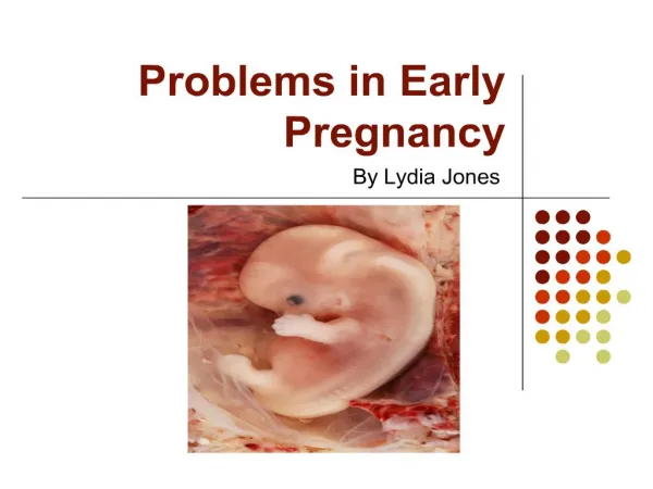
Definition Termination of pregnancy before viability of the foetus i.e. before 28 weeks (in Britain) and before 20 weeks or if the foetal weight is less than 500 gm (in USA and Australia). When the abortion occurs spontaneously, the term " miscarriage" is often used.
Aetiology • Chromosomal abnormalities: cause at least 50% of early abortions e.g. trisomy. • Blighted ovum (anembryonic gestational sac). • Maternal infections: Acute fever for whatever the cause can induce abortion. • Trauma: external to the abdomen or during abdominal or pelvic operations. • Endocrine causes: Progesterone deficiency ,Diabetes mellitus, Hyperthyroidism. • Drugs and environmental causes: • Maternal anoxia and malnutrition. • Over distension of the uterus: e.g. acute hydramnios.
Continued, • Immunological causes: • Systemic lupus erythematosus. • Antiphospholipid antibodies that are directed against platelets and vascular endothelium leading to thrombosis, placental destruction and abortion. • Ageing sperm or ovum. • Uterine defects Septum, Asherman's syndrome (intrauterine adhesions). • Nervous, psychological conditions and over fatigue. • Idiopathic.
Threatened Abortion Clinical picture: • Symptoms and signs of pregnancy coincide with its duration. • Vaginal bleeding slight or mild, bright red in colour. • Pain is absent or slight. • Cervix is closed. • Pregnancy test is positive. • Ultra-sonography shows a living foetus.
Prognosis: • If the blood loss is less than a normal menstrual flow and is not accompanied by pain of uterine contraction there is a reasonable chance for continuing pregnancy. This occurs in 50% of cases while other half will proceed to inevitable or missed abortion.
Treatment: • Rest in bed until one week after stoppage of bleeding. • No intercourse as it may disturb pregnancy by the mechanical effect and the effect of semen prostaglandins on the uterus. • Sedatives: if the patient is anxious. • Treatment of controversy: • Progestogens. • Gonadotrophins may be of benefit in cases of luteal phase deficiency and those get pregnant with ovulatory drugs.
Inevitable Abortion Clinical picture: • Symptoms and signs of pregnancy coincide (match) with its duration. • Vaginal bleeding is excessive and may accompanied with clots. • Pain is colicky felt in the suprapubic region radiating to the back. • The internal os of the cervix is dilated and products of conception may be felt through it. • Rupture of membranes between 12-28 weeks is a sign of the inevitability of abortion.
Treatment • Any attempt to maintain pregnancy is useless.
Incomplete Abortion • Retention of a part of the products of conception inside the uterus. It may be the whole or part of the placenta which is retained.
Clinical picture • The patient usually noticed the passage of a part of the conception products. • Bleeding is continuous. • The uterus is less than the period of amenorrhoea but still large in size. The cervix is opened and retained contents may be felt through it. • Ultrasonography: shows the retained contents.
Complete Abortion • All products of conception have been expelled from the uterus. Clinical picture: • The bleeding is slight and gradually diminishes. • The pain ceases. • The cervix is closed. • The uterus is slightly larger than normal. • Ultrasound: shows empty cavity.
Missed Abortion • Retention of dead products of conception for 4 weeks or more. Symptoms: • Symptoms of threatened abortion may or may not be developed. • Regression of pregnancy symptoms as nausea, vomiting and breast symptoms. • The abdomen does not increase and may even decrease in size. • The foetal movements are not felt or ceases if previously present. • A dark brown vaginal discharge may occur (prune juice discharge).
Signs: • The uterus fails to grow and becomes firmer and The cervix is closed. • The foetal heart sounds cannot be heard. Investigations: • Pregnancy test becomes negative within two weeks from the ovum death. • Ultrasound shows either a collapsed gestational sac, absent foetal heart movement or foetal movement.
Complications: • Disseminated intravascular coagulation (DIC) may occur if the dead conceptus is retained for more than 4 weeks. • Superadded infection.
Treatment: • The dead conceptus is expelled spontaneously in the majority of cases. Evacuation of the uterus is indicated in the following conditions: • spontaneous expulsion does not occur within four weeks, • there is bleeding, • infection or DIC developed or, • patient is anxious. Although some gynaecologists advise evacuation of the uterus once sure diagnosis of missed abortion is made.
Evacuation is carried out as following: • If the uterine size is less than 12 weeks’ gestation: vaginal or suction evacuation is done • If the uterine size is more than 12 weeks' gestation: evacuation can be done by • Prostaglandins: given intravaginally (PGE2), intravenously, intra-or extra- amniotic (PGF2α). • Oxytocin infusion. • Combination. • Hysterotomy: is rarely indicated in 2nd trimester missed abortion if the medical induction fails initially and after repetition few days later.
Septic Abortion • It is any type of abortion, usually criminal abortion, complicated by infection. • Microbiology: • E.Coli, bacteroids, anaerobic streptococci, clostridia, streptococci and staphylococci are among the most causative organisms.
Clinical picture: • General examination: • Pyrexia and tachycardia. • Rigors suggest bacteraemia. • A subnormal temperature with tachycardia is ominous and mostly seen with gas forming organisms. • Malaise, sweating, headache, and joint pain. • Jaundice and /or haematuria is an ominous sign, indicating haemolysis due to chemicals used in criminal abortion or haemolytic infection as clostridium welchii.
Abdominal examination: • Suprapubic pain and tenderness. • Abdominal rigidity and distension indicates peritonitis. • Local examination: • Offensive vaginal discharge. Minimal inoffensive vaginal discharge is often associated with severe cases. • Uterus is tender. • Products of conception may be felt. • Local trauma may be detected. • Fullness and tenderness of Douglas pouch indicates pelvic abscess which will be associated with diarrhoea.
Treatment • Isolate the patient . Bed rest in semi-sitting position??????????????????. • An intravenous line is established for therapy. • Observation for vital signs: • A cervico-vaginal swab is taken for culture and sensitivity, • Antibiotic therapy:. • Fluid therapy: • Blood transfusion: is given if CVP is low (normal: 8-12 cm water).
Continued, • Oxytocin infusion: to control bleeding and enhances expulsion of the retained products. • Surgical evacuation of the uterus can be done after 6 hours of commencing IV therapy but may be earlier in case of severe bleeding or deteriorating condition in spite of the previous therapy. • Hysterectomy may be the last choice to safe life
Other types of abortion Therapeutic Abortion • Abortion induced for a medical indication. Criminal Abortion • Illegal abortion induced for a non-medical indication. Recurrent (Habitual) Abortion • Three (two by some authors) or more consecutive abortions.
Definition • Ectopic means "out of place." In an ectopic pregnancy, a fertilized egg has implanted outside the uterus. The egg settles in the fallopian tubes in more than 95% of ectopic pregnancies. This is why ectopic pregnancies are commonly called "tubal pregnancies.
Signs and Symptoms • Ectopic pregnancy can be difficult to diagnose because symptoms often mirror those of a normal early pregnancy. These can include missed periods, breast tenderness, nausea, vomiting, or frequent urination. • The first warning signs of an ectopic pregnancy are often pain or vaginal bleeding.
Fate of ectopic pregnancy • Tubal abortion • Tubal rupture
Morbidity and Mortality Rates • Abdominal pain occurs in 97% of women with an ectopic pregnancy, • Vaginal bleeding in 79%, • abdominal tenderness in 91%, and infertility in 15%. • Persistent ectopic pregnancy after surgical treatment occurs in 5–10% of cases. • Ectopic pregnancy accounts for 10–15% of all maternal death; the mortality rate for ectopic pregnancy is approximately one in 2,500 cases.
Hydatidiform Mole • A hydatidiform mole is a relatively rare condition in which tissue around a fertilized egg that normally would have developed into the placenta instead develops as an abnormal cluster of cells. (This is also called a molar pregnancy.) This grapelike mass forms inside of the uterus after fertilization instead of a normal embryo.
Causes • The cause of hydatidiform mole is unclear; • some experts believe it is caused by problems with the chromosomes • A mole sometimes can develop from placental tissue that is left behind in the uterus after a miscarriage or childbirth.
Symptoms • Women with a hydatidiform mole will have a positive pregnancy test and often believe they have a normal pregnancy for the first three or four months. • However, in these cases the uterus will grow abnormally fast. • By the end of the third month, if not earlier, the woman will experience vaginal bleeding ranging from scant spotting to excessive bleeding. • Sometimes, the grapelike cluster of cells itself will be shed with the blood during this time. • Other symptoms may include severe nausea and vomiting and high blood pressure. As the pregnancy progresses, the fetus will not move and there will be no fetal heartbeat.
Prognosis • A woman with a molar pregnancy often goes through the same emotions and sense of loss. • In addition, there is the added worry that the tissue left behind could become cancerous. • In the unlikely case that the mole is cancerous the cure rate is almost 100%. As long as the uterus was not removed, it would still be possible to have a child at a later time.