Download
1 / 27
280 likes | 521 Vues
Non-Invasive Imaging of Protein Interactions within Living Models. Sean Courtney. Why study Protein Interactions?. Regulates many of the essential biological processes: Transcription Translation Metabolic pathways Signal Transduction Cell Cycle Progression etc.

E N D
Non-Invasive Imaging of Protein Interactions within Living Models Sean Courtney
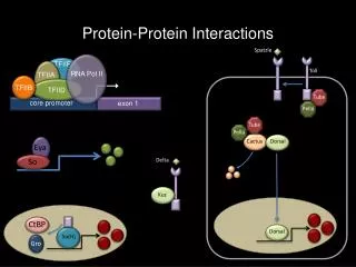
Why study Protein Interactions? • Regulates many of the essential biological processes: • Transcription • Translation • Metabolic pathways • Signal Transduction • Cell Cycle Progression • etc. • Yields information on possible roles of genes with as of yet unknown functions • Detects novel interactions between proteins of other various functions http://www.wellesley.edu/Chemistry/chem227/nucleicfunction/transcription/lac%20operon/06eukaryotes.jpg
Protein-Protein Interaction Techniques • Split Ubiquitin • Protein Fragment Complementation • Yeast Two-Hybrid http://www.ittc.ku.edu/~xwchen/Project_files/image007.jpg http://www.wesleyan.edu/mbb/faculty/imukerji/hbs3.jpg
Split Ubiquitin • Fusions of Ubiquitin (Ub) and a target protein are recognized and cleaved by Ub-specific Proteases (UBPs), that recognize the folded conformation of Ub not the sequence • Ub can be expressed in yeast as an N-terminal half (Nub) and a C-terminal half (Cub), which have affinity for each other and spontaneously assemble forming the “split ubiquitin” • If Nub and Cub are coexpressed in a single cell, the reporter protein will be cleaved upon reassembly • The bait protein is fused to Cub followed by a reporter protein and a prey protein is fused to a mutated Nub (NubG, lost affinity) • If interaction occurs then the NubG and Cub are brought into proximity allowing reassembly and cleavage by the UBPs, thus releasing the reporter protein Dualsystems Biotech
Protein Fragment Complementation • Uses an enzyme that usually produces a detectable product (colorimetric, fluorometric, or survival) • i.e. murine dihydrofolate reductase (mDHFR) – reduces dihydrofolate into tetrahydrofolate • A bait protein is fused to one “part” of the enzyme and a prey protein is fused to the other “part” of the enzyme, both of which are transfected/transformed into cells • If the proteins interact, they bring the subunits of the enzyme within a close proximity, thus enabling their reassembly into an active enzyme, upon which the enzyme’s substrate is added and the product can then be detected Michnick et al. 1998
Yeast Two-Hybrid • Gal4 is a yeast transcription factor • Each Gal4-responsive gene contains a target sequence, UAS • When Gal4 binds the UAS, transcription is activated from a downstream promoter • Bait gene fused to a GAL4 DNA-BD and a cDNA library (or another known protein) fused to a GAL4 DNA-AD • GAL4 DNA-BD can bind the UAS alone but cannot activate transcription until bound with the GAL4 DNA-AD • When the two domains interact, BD and AD are brought into proximity, thus activating transcription of a downstream reporter gene Clontech
Various Imaging Techniques • Magnetic Resonance Imaging (MRI) • Positron Emission Tomography (PET) • Single-Photon Emission Computed Tomography (SPECT) • Fluorescence Resonance Energy Transfer (FRET) • Fluorescence • Bioluminescence
Magnetic Resonance Imaging (MRI) • Used to visualize the inside of living organisms • Demonstrates pathological or other physiological alterations of living tissues (i.e. tumors) • Uses radio frequency signals to acquire images • Based on the relaxation properties of excited Hydrogen nuclei in water http://en.wikipedia.org/wiki/Image:User-FastFission-brain.gif http://en.wikipedia.org/wiki/Image:3Dbrain.gif
MR Imaging Ability • http://video.google.com/videoplay?docid=3477454458695092843&q=MRI&hl=en
Positron Emission Tomography (PET) • A nuclear medicine imaging technique that produces a 3D image or map of functional processes in the body • Uses a short-lived radioactive tracer isotope which decays by emitting a positron (has been chemically incorporated into a metabolically active molecule) and is injected into the living animal, usually in the blood • A waiting period ensues while the metabolically active molecule (usually fluorodeoxyglucose, FDG) becomes concentrated in tissues of interest • Result of two simultaneous annihilation photons emitted back-to-back • The image produced is not the location of the radionucleoside but that of where the annihilation event occurs • Commonly used alongside CT scans or MRI scans, giving both anatomic and metabolic information http://en.wikipedia.org/wiki/Image:PET-MIPS-anim.gif
Single-Photon Emission Computed Tomography (SPECT) • A nuclear medicine tomographic imaging technique using gamma rays able to provide true 3D information • A 2D view of the 3D distribution of a radionucleotide from multiple angles • A computer is used to apply a tomographic reconstruction algorithm to yield a 3D dataset • Can be manipulated to show thin slices along any chosen axis of the body Fluorescence Resonance Energy Transfer (FRET) • Energy transfer mechanism between two fluorescent molecules • Useful tool to quantify molecular dynamics in biophysics, such as protein-protein interactions, protein-DNA interactions, and protein conformational changes • Monitors the complex formation between two molecules, one is labeled with a donor and the other with an acceptor, which are then mixed • When they dissociate, the donor emission is detected upon the donor excitation, but when together, the acceptor emission is predominant
Fluorescence • Production and emission of light by a living organism as the result of a chemical reaction during which chemical energy is converted to light energy • Uses an external light source with a low-pass filter to excite the fluorescent molecules • Green Fluorescent Protein, originally found in the Aequorea victoria species of jelly fish • Been biochemically modified to produce Green, Yellow, Blue, Cyan, and Red Fluorescent Proteins for use in various research techniques using a reporter • Limited by tissue autofluorescence, as well as the light being able to first get into the living model and sensing the target fluorescent molecule, then having that fluorescence get back out of the model and to the detector (a lot of scattering occurs) http://en.wikipedia.org/wiki/Image:Aequorea_victoria.jpg http://wwwchem.leidenuniv.nl/metprot/armand/images/029l.jpg http://www.upenn.edu/pennnews/photos/704/mice.jpg
Bioluminescence • Luciferase, an enzyme found in Fireflies, is also commonly used as a reporter • Must be incorporated into the cell (i.e. tumor xenograft) • Just before imaging, luciferin is added via IV or IP, which can then rapidly travel throughout the body and where ever it encounters luciferase, oxygen, and ATP it will be converted to oxyluciferin and produce a detectable light • Pros: No external light source, no autofluorescent background noise from surrounding tissues, and depth of penetration is not as limiting compared to its fluorescent counterparts • Cons: Limited to studying genetically modified cells, transgenic animal models, or infectious agents • Gives a very weak signal and requires a highly efficient Charge-coupled device (CCD) because it has a strong dependence of signal intensity on source depth Cherry et al. 2004
How to use the Molecular Imaging Techniques to View Protein Interactions in Living Models • Must first define your target of interest usually Cell-Surface Receptors or Enzymes • Once chosen, need to next choose the method to view it in the living model and therefore need to also choose the contrast agent • Dependent on spatial arrangement and the desired resolution, location and distribution of the target, concentration of the target, and the specificity of the target • Need an exogenous agent detectable by its physical or chemical properties • Common agents include radioactive atoms, fluorescent molecules, paramagnetic ions, or small molecules covalently linked to the target with similar properties as those listed
Various Contrast Agents Cherry et al. 2004
Cell-Surface Receptor • Similar concept to pharmaceuticals in that it must find the target in the body and accumulate there • Binds to the target, unbound portion must be cleared from that tissue to be able to distinguish the signal between specific and non-specific • Administration is usually intravenously (IV) into the bloodstream where it can rapidly travel through the body, sometimes injection is intraperitoneal (IP) within the abdominal cavity • The target must successfully “trap” the signal molecule within the cell or tissue and thus accumulate to provide an adequate signal Cherry et al. 2004
Enzymes • Agents are designed to interact with the enzyme target • Interaction of the signal agent with the enzyme causes a change in the agent (i.e. charge) so that it remains in that cell, “trapped” • Gadolinium (Gd3+) • Activatable agent • Highly paramagnetic • Enclosed in a molecular “case” where it is unable to interact with water in the tissue • Interaction with the target enzyme breaks the linker (the “lid”) thus causing a structural change in the molecule where the Gd3+ can now interact with the water, changing the relaxation state Cherry et al. 2004
Reporter Gene Method of Detection • Genetically modify the cell • Place the reporter gene under the control of the same promoter of the gene of interest • cells expressing the reporter also express the gene of interest in a ratio of approximately 1:1 • This is limiting because the reporter must be introduced into the living model • Introduce the reporter gene into cancer cell lines to track cancer cells and their progeny via xenographs and tumor transplant models Cherry et al. 2004
Various Reporter Gene Systems Cherry et al. 2004
“Noninvasive imaging of protein-protein interactions in living animals” • Gary D. Luker, Vijay Sharma, Christina M. Pica, Julie L. Dahlheimer, Wei Li, Joseph Ochesky, Christine E. Ryan, Helen Piwnica-Worms, and David Piwnica-Worms • Molecular Imaging Center, Mallinckrodt Institute of Radiology and Departments of Molecular Biology and Pharmacology, Cell Biology and Physiology, and Internal Medicine, and Howard Hughes Medical Institute, Washington University School of Medicine, St. Louis, MO 63110 • Proposal: To develop a method for detecting protein-protein interactions in living mice by combining the yeast two-hybrid system with various reporter proteins sufficient for imaging
Construction of Reporter Proteins • HSV-1-TK Nucleoside analogs are actively transported into cells and are preferentially phosphorylated by the viral TK and not the mammalian TK • Previous mutagenesis studies by Black et al. showed a mutant HSV-1-TK with enhanced sensitivity to 8-18F-fluoropenciclovir (PCV) HSV-1-sr39TK • Degreve et al. showed that a mutation in the NLS sequences of HSV-1-TK provided better uptake of 124I-FIAU • Disrupted one N-terminal NLS sequence of the HSV-1-TK NLS mNLS-sr39TK • Fused EGFP into both • HSV-1-sr39TK-EGFP • mNLS-sr39TK-EGFP
Characterization of the Two-Hybrid System • Treatment with Doxycycline activates a reverse tetracycline-responsive transactivator inducing bi-directional transcription • p53 and TAg are known to interact • BD binds the promoter and upon interaction between p53 and TAg, AD is now in place to promote transcription of the downstream reporters • mNLS-sr39TK can be used for microPET • EGFP can be used for Fluorescence Microscopy
Development of a Reporter Cell Line • Stably transfected HeLa cells with Gal4-mNLS-sr39TK-EGFP to develop a reporter cell lineHeLa-Gal4 • Only cells expressing p53 and TAg showed cellular accumulation of PCV • Stably transfected HeLa-Gal4 cells with the various constructs and treated with doxycycline • Only expressed upon antibiotic treatmenttightly regulated expression • Measured the activity of the nucleoside analog reporter • Only saw activity when cells expressed both p53 and TAg, with treatment • GFP expression only in the presence of doxycycline
Time course of Reporter Gene Induction • To determine the peak expression time of the reporter, cells either expressing p53/CP or p53/TAg were treated with doxycycline for the displayed times and PCV accumulation was then measured • Peak expression time was at 48 h and then began to decrease thereafter
Imaging in Vivo Protein Interactions • Produced xenograft tumors of TAg and CP cells of nude mice • Once tumors grew to ~5 mm, mice were treated and imaged 1h after tail injection of 18F-FHGB • Fluorescent microscopy of excised tumors displayed expression of GFP • Treated TAg tumor mice with doxycycline for 12, 24, and 48 h to determine if microPET could quantify hybrid protein levels • Also looked at the biodistribution of 18F-FHGB • The intensity of the reporter was proportional to the amount of interacting proteins in vivo
Conclusions • Binding of p53/TAg in living mice was detected by microPET imaging with 18F-FHGB • An approximate 6-fold increase in mNLS-sr39TK activity occurred in response to the interaction of p53 and TAg in vivo • Function of the reporter protein was enhanced when increasing the amounts of interacting proteins were used • In vivo microPET imaging could be used to determine relative affinity differences between interacting proteins previously shown in vitro • GFP fluorescent imaging in vivo could provide a rapid screening assay for detecting the presence or absence of protein-protein interactions • Xenograft models may be useful in the initial characterization of drugs targeted to specific protein interactions • Transgenic mice with reporter genes could allow the interaction of proteins to be monitored in their native environment • Potential to advance our understanding of how protein interactions affect our normal physiology, development, disease progression and response to therapy
References • Cherry, S. R. (2004). "In vivo molecular and genomic imaging: new challenges for imaging physics." Phys Med Biol49(3): R13-48. • Gross, S. and D. Piwnica-Worms (2005). "Spying on cancer: molecular imaging in vivo with genetically encoded reporters." Cancer Cell7(1): 5-15. • Gross, S. and D. Piwnica-Worms (2006). "Molecular imaging strategies for drug discovery and development." Curr Opin Chem Biol10(4): 334-42. • Haberkorn, U. and A. Altmann (2003). "Noninvasive imaging of protein-protein interactions in living organisms." Trends Biotechnol21(6): 241-3. • Luker, G. D., J. P. Bardill, et al. (2002). "Noninvasive bioluminescence imaging of herpes simplex virus type 1 infection and therapy in living mice." J Virol76(23): 12149-61. • Luker, G. D., V. Sharma, et al. (2002). "Noninvasive imaging of protein-protein interactions in living animals." Proc Natl Acad Sci U S A99(10): 6961-6. • Luker, G. D., V. Sharma, et al. (2003). "Molecular imaging of protein-protein interactions: controlled expression of p53 and large T-antigen fusion proteins in vivo." Cancer Res63(8): 1780-8. • Luker, G. D., V. Sharma, et al. (2003). "Visualizing protein-protein interactions in living animals." Methods29(1): 110-22. • Luker, K. E., M. C. Smith, et al. (2004). "Kinetics of regulated protein-protein interactions revealed with firefly luciferase complementation imaging in cells and living animals." Proc Natl Acad Sci U S A101(33): 12288-93. • Winnard, P., Jr. and V. Raman (2003). "Real time non-invasive imaging of receptor- ligand interactions in vivo." J Cell Biochem90(3): 454-63.