
Uploaded by
griffith-ward
1 SLIDES
129 VUES
10LIKES
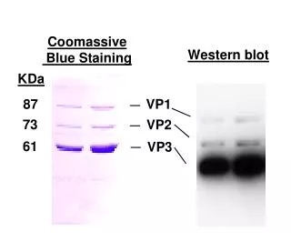
Coomassie Blue Staining for Western Blot Analysis of Viral Proteins VP1, VP2, and VP3
DESCRIPTION
This study explores the application of Coomassie Blue staining in Western blotting to visualize viral proteins, specifically VP1 (87 kDa), VP2 (73 kDa), and VP3 (61 kDa). The methodology emphasizes the effectiveness of Coomassie Blue for protein detection, offering insights into the molecular weight and expression levels of these important viral components. This approach enhances understanding of viral pathogenesis and aids in the development of diagnostic tools and therapeutic strategies.
Download
1 / 1
Télécharger la présentation 
Coomassie Blue Staining for Western Blot Analysis of Viral Proteins VP1, VP2, and VP3
An Image/Link below is provided (as is) to download presentation
Download Policy: Content on the Website is provided to you AS IS for your information and personal use and may not be sold / licensed / shared on other websites without getting consent from its author.
Content is provided to you AS IS for your information and personal use only.
Download presentation by click this link.
While downloading, if for some reason you are not able to download a presentation, the publisher may have deleted the file from their server.
During download, if you can't get a presentation, the file might be deleted by the publisher.
E N D


More Related