Download
1 / 28
320 likes | 475 Vues
This article explores various types of refraction, including emmetropy, myopia, hypermetropy, and astigmatism. It covers the clinical classification of myopia by degree and genesis, discussing uncomplicated, inherited, and complicated forms. The degrees of myopia and hyperopia are detailed alongside their associated complications. Additionally, the piece reviews LASIK techniques for correcting refractive errors and outlines the benefits and drawbacks of alternative surgical methods, emphasizing advancements in laser ophthalmology. Gain insights into effective vision restoration strategies.

E N D
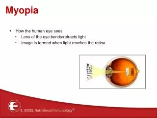
Types of refraction 1.Emmetropy 2. Myopia 3. Hypermetropy 4. Astigmatism
Clinical classification of myopia By degree By genesis By type of disease By presence of complications Low Uncomplicated Inherited Stationary Slowly prosgressive Complicated Congenital Middle Quickly progressive Acquired High
Degrees of myopia • Myopia of low degree • Under (-) 3.0 diopters • Myopia of moderate degree • From (-) 3.0 to (-) 6.0 diopters • Myopia of high degree • Higher than (-) 6.0 diopters
Degrees of hyperopia • Hyperopia of low degree • Under (+)2 diopters • Hyperopia of moderate degree • From (+)2 to (+) 5 diopters • Hyperopia of high degree • Higher than (+) 5 diopters
Complications of hypermetropy Sty, Conjunctivitis, Blefaritis. Concomitant convergent strabismus Accomodative astenopy Pseudoneuritis
Myopic cone Posterior staphyloma in the beginning myopia
Scimitar tear of ocular choroid Tear of ocular choroid
Druses of vitreous lamina Tear of ocular choroid
True staphyloma of sclera at myopia of high degree Choroiditis at myopia
Ocular fundus at myopia of high degree Ocular fundus at myopia of high degree
Hemorrhage in the macula at myopia of high degree Macular pigmentation in place of older recurrent hemorrhages at myopia of high degree
ILLUSTRATED REVIEW OF LASIK TECHNIQUE With the help of mechanic instrument – microkeratom – a round flap is opened in the protective layer of cornea.
ILLUSTRATED REVIEW OF LASIK TECHNIQUE Upper layers of corneal stroma are evaporated by eximer laser on the necessary for accurate focusing depth.
ILLUSTRATED REVIEW OF LASIK TECHNIQUE In the result of operation the form of cornea became planer. Saved natural layer provides quick rehabilitation of patient.
Refractive errors correction • SPECTACLES AND CONTACT LENSES • SURGICAL INTERVENTION • 3. LASER CORRECTION
SHORT VISION RECOVERY PERIOD POSSIBILITY TO MAKE SURGERY ON BOTH EYES SIMULTANEOUSLY STABLE POSITIVE RESULT LASIC ADVANTAGES
RADIAL KERATOTOMY ADVANTAGES: • POSSIBILITY TO CORRECT MYOPIA UP TO 5 DIOPTERS DISADVANTAGES: • LONG REHABILITATION PERIOD • RISK OF DEVELOPMENT OF WRONG ASTIGMATISM
LASER OPHTHALMOLOGY Many scientists made attempts to use light beam in ocular diseases. The first to realize this idea was Myer-Schwickerath in 1947. This date may be considered as the beginning of clinical use of photocoagulation in ophthalmology. Meyer-Schwickerath used xenon lamp of high pressur as a powerful source of light.