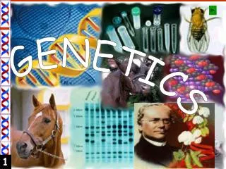
Genetics
Genetics. Instructor: Dr. Jihad Abdallah Lecture 2 The cell cycle and Cell Division. The cell cycle. Living cells go through a series of stages known as the cell cycle. They undergo a continuous alternation between division and non-division.

Genetics
E N D
Presentation Transcript
Genetics Instructor: Dr. Jihad Abdallah Lecture 2 The cell cycle and Cell Division
The cell cycle • Living cells go through a series of stages known as the cell cycle. They undergo a continuous alternation between division and non-division. • The period between cell divisions is known as interphase. Interphase consists of three phases (G1 phase, S phase, and G2 phase). • During interphase, the biochemical activity is devoted to : - Cell growth - Replication of DNA of each chromosome
- Interpahse: 1 - G1 (Gap 1): - In this phase the cell grows. - Synthesis of enzymes necessary in the S phase. - At some point in G1 the cell follows one of two paths: either inter a resting phase called G0 stage (Gap 0) or become committed to initiate DNA synthesis and complete the cycle. The time when this decision is made is called G1 checkpoint 2- S phase: The period during which DNA is synthesized. The cell makes copies of its chromosomes. Each chromosome now consists of two sister chromatids. 3 - G2 (Gap 2): - Second growth phase. - The cell checks the duplicated chromosomes and gets ready to divide. - By the end of G2 the volume of the cell has roughly doubled, DNA has been replicated and mitosis is initiated. - M phase (Mitosis): the cell divides into two new cells.
Duration of the cell cycle in Human cells (in hours) It takes about 16 hours Duration of phases of Mitosis: Prophase: 36 minutes Metaphase: 3 minutes Anaphase: 3 minutes Telophase: 18 minutes Vary among cell types Consistent among cell types
Mitosis • Mitosis consists of the following stages: • Prophase: - the centrioles migrate to two opposite sides of the cell. - spindle fibers start to form - the nuclear envelope begins to break down and the nucleolus fades. - Microtubules of the cytoskeleton disassemble. - The diffuse chromatin (replicated DNA and associated proteins) condenses into chromosomes. - Each replicated chromosome comprises two chromatids, both with the same genetic information (called sister chromatids).
2.Prometaphase: - In this stage the nuclear envelope breaks down so there is no longer a recognizable nucleus. - Some mitotic spindle fibers (microtubules) elongate from the centrosomes and attach to kinetochores (protein bundles at the centromere region on the chromosomes where sister chromatids are joined). - These microtubules which attach to the kinetochore are called “kinetochore microtubules”. - Other spindle fibers elongate but instead of attaching to kinetochores, they attach with spindle fibers growing from the other side of the cell. These are called “polar microtubules or non-kinetochore microtubules”. - the chromosomes start to migrate towards the center of the cell by the kinetochore microtubules
3. Metaphase: all chromosomes align in one plane at the center of the cell called the equatorial plane (also referred to as the metaphase plate). 4.Anaphase: Spindle fibers shorten, the kinetochores separate, and the sister chromatids (daughter chromosomes) are pulled apart and begin moving to the cell poles. 5. Telophase : - The daughter chromosomes arrive at the poles and the spindle fibers that have pulled them apart disappear. - Cytokinesis of the cytoplasm (division of the cytoplasm) occurs resulting in two identical cells. - In each new cell, chromosomes begin to uncoil and become diffuse chromatin while a nuclear envelope re-forms arround them. - The nucleolus gradually re-forms
Meiosis (reduction division) • Meiosis produces gametes with only one haploid set of chromosomes (reduction of the number of chromosomes by half) • Meiosis is divided into two main steps: • Meiosis I • Meiosis II
Meiosis I • Prophase I divided into 5 substages: • Leptonema (Leptotene stage) • Zygonema (Zygotene stage) • Pachynema (Pachytene stage) • Diplonema (Diplotene stage) • Diakinesis • Metaphase I • Anaphase I • Telophase I
Prophase 1 • Leptonema: (from Greek words meaning "thin threads“) - Chromtin material begins to condense and the chromosomes become visible • Each chromosome begins to search its homologue (homology search)
Zygonema: "paired threads" - The chromosomes continue to condense • Homologous chromosomes find each other and begin to align to each other in a process known as “rough pairing” • A synaptonemal complex starts to form (synapsis begins) between the homologs. • At the completion of zygonema the paired homologs represent structures referred to as “bivalents” (also called tetrads because each one consists of four chromatids two sister chromatids and two non-sister chromatids)
In pachynema: "thick threads" • The aligned homologous chromosomes become much more closely associated. This process is known as synapsis. (The chromosomes are said to have synapsed) • The chromosomes continue to condense. • Crossing-overoccurs(Exchange of genetic material between non-sister chromatids of homologous chromosomes) between non-sister chromatids but the result of crossing-over is only visible when the chromosomes begin to separate.
Diplonema: "two threads" • The homologous chromosomes in each tetrad begin to separate, but they remain connected at points of crossing over. Each point of crossing over is known as a chiasma (plural: chiasmata). • Also at this stage, the nuclear envelope begins to break down.
Diakinesis:"moving through" • - Is the last stage of prophase I. • The chiasmata proceed to the end of the chromatids, then separate (terminalization). This leaves chromatids that engaged in crossing over with exchanged genetic material • The nucleolus and nuclear envelope break down. • The centromeres of the chromosomes become attached to spindle fibers.
Metaphase I • Chromosomes (bivalents with each one consisting of two chromatids) align at the center of the cell (the metaphase plate) • A single centromere holds each pair of sister chromatids together. • The centromere does not divide and the two sister chromatids remain attached (Unlike mitosis where the centromere divides in Anaphase)
Anaphase I • One-half of each tetrad (one pair of sister chromatids) called a “dyad” is pulled towards one pole of the cell at random. • This process is called disjunction
Telophase I • Chromosomes (each consisting of two chromatids) complete their migration to the poles. • A nuclear membrane forms around each set of dyads. • Cytokinesis occurs during Telophase I so that two cells are produced each containing half the number of dyads.
Meiosis II (Similar to mitosis) • Prophase II: - each dyad is composed of one pair of sister chromatids attached by one centromere - the nuclear membrane that formed during Telophase I breakdown and chromosomes recondense • Metaphase II: - the dyads move and align at the center of the cell - then the centromere divide • Anaphase II: - the sister chromatids of each dyad separate and begin to move towards the opposite poles of each cell - each chromatid is now considered a separate chromosome called “monad” • Telophase II: - nuclear membranes form again and Cytokinesis occurs - at the end of Meiosis II, 4 haploid cells are produced from a single cell entering meiosis.