Download
1 / 41
620 likes | 2.61k Vues
Injuries of the Fingertip. presented by Spencer F. Schuenman, D.O. Distal Fingertip Injuries. Amputations Injuries of the Perionychium Infections. The Goals of Amputation Surgery. 1. Preservation of functional length 2. Preservation of useful sensibility

E N D
Injuries of the Fingertip presented by Spencer F. Schuenman, D.O.
Distal Fingertip Injuries • Amputations • Injuries of the Perionychium • Infections
The Goals of Amputation Surgery • 1. Preservation of functional length • 2. Preservation of useful sensibility • 3. Prevention of symptomatic neuromas • 4. Short morbidity • 5. Early return to work or play
Amputations with Skin or Pulp loss only • Treatment options include: • split thickness skin grafting • primary closure • healing by secondary intention • V-Y skin flaps • Complications include: reduced sensibility, cold sensitivity, tenderness, infection.
Amputations with Exposed Bone • Local Flap Coverage • Atasoy-Kleinert Volar V-Y Flap • Kutler’s Lateral V-Y Flaps • Volar Flap Advancement • Cross-Finger Pedicle Flap • Thenar Flap • Thenar H-Flap (Smith and Albin)
Summary of Treatment Options for Amputations • All of the studies on the various techniques reported far superior results by the surgeons that devised the particular procedure that was named after them. • From comparison evaluations of the literature it was apparent that distal tip amputations without exposed bone can be treated nonoperatively with good results (aprox. 90%).
Distal tip amputations with exposed bone can be converted to one without exposed bone by ronguering the protruding portion of the phalanx and treating it conservatively.
If surgical intervention is chosen, it has been shown that all of the aforementioned techniques can be equally successful, with comparable complications, so it is prudent to perfom the procedure that is most familiar to the surgeon.
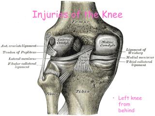
Perionychial Injuries • Anatomy- consists of the paronychium and the nail bed • The proximal nail fits into a depression called the nail fold, the skin over the dorsum of the nail fold is the nail wall. The thin membrane extending from the nail wall onto the the dorsum of the nail is the eponychium. The lunula is the curved, white opracity in the nail found just distal to the eponychium and is at the junction of the germinal and sterile matrixes.
The nail bed is all the soft tissue immediately beneath the nail that participates in nail generation and migration. The mass of keratin between the distal nail and the nail bed is the hyponychium. This part is very resistant to infection • The nail bed consists of the germinal and sterile matrixes.
Physiology-complete longitudinal nail growth takes between 70 and 160 days at a rate of approximately 0.1 mm/day. • Fingernails grow more rapidly than do toenails by a ratio of 4 : 1. • Ridges are scars in the nail bed. • Shininess of the nail is produced by an intact dorsal roof and if damaged dullnes results.
Treatment of Acute Injuries • Subungal Hematomas-use of a drill, needle, or paper clip heated until red hot and then making a hole in the nail at the site of the hematoma
Simple Lacerations-most common-remove the nail with a small periosteal elevator and then clean and soak the nail in a betadine solution. Then explore the nail bed and determine the amount of crush and irregularities of the edges. Trim the irregularities. Then approximate the edges with a 5,6,or 7-0 chromic suture. The nail is then reinserted into the nail fold and can be sutured into place if desired.
Stellate Lacerations-The etiology of stellate lacerations is similar to simple. Accurate, meticulous approximation of the stellate points is necessary to achieve a good result.
Severe crush-These injuries carry a poorer prognosis. It is necessary that all fragments of the nail bed be returned to the nail bed and repaired as accurately as possible. • Lacerations associated with fractures of the distal phalanx-approx. 50% of nail bed injuries are associated with a fracture. The fracture needs to be reduced and either splinted by the nail itself or with fine K-wires.
Avulsion-frequently leaves the fragment of nail bed attached to the undersurface of the avulsed nail. An attempt should be made to find the nail because the nail to which the bed is attached is the optimum shape and tension to aid inosculation of the nail bed. The nail should, therefore, be replaced as accurately as possible without separating the nail from the bed.
Summary • Nail bed injuries should be repaired as soon as possible • The nail should not be removed from the nail bed if at all possible • All of the margins of the nail bed should be meticulously repaired • The associated fracture should be properly reduced and fixated with the use of K-wires if necessary
Infections of the Fingertip • General Principles-If the problem is diagnosed within 48 hours of onset, high doses of systemic antibiotics can arrest the problem. Beyond this, success decreases because of thrombosis of the local small vessels in many areas of the hand. Many closed spaces and compartments in the hand permit early and rapid walling off of any infection, thus preventing antibiotics from reaching the infected area.
The most common infecting organism is Staphylococcus aureus, so the choice of agent should be based on this. • Antibiotics should initially be given intravenously for 2-3 days, then they can be given orally for a total of aprox. 7-10 days. It is not necessary to give prolonged use of Abxs in soft tissue infections.
Infections usually begin as a cellulitis, if an abscess forms incision and drainage is necessary. All incisions should permit easy extension in any direction in case the magnitude of the problem is greater than anticipated. Correlatively, the incision or its extension should not traverse any flexion crease at or approaching a right angle, lest a flexion contracture develop.
The approach should avoid injuring any vessels, nerves, or tendons. All procedures should be carried out with the use of tourniquet, careful not to exanguinate to forcefully as to not squeeze the bacteria into the bloodstream and seed other areas of the body.
In other techniques the incisions are left open and are allowed to close by secondary intention. The wounds should be packed for 48-72 hours, followed by saline soaks and exercises.
Various Infections • Paronychia • Chronic Paronychia • Felons • Pyogenic Flexor Tenosynovitis
Paronychia • This infection involves the soft tissue fold around the fingernail. It usually results from introduction of S. aureus into the tissue by a sliver of nail or hangnail, a manicure instrument, or a tooth. • Paronychia are the most common infection of the hand.
Treatment • early stages-the process can be aborted by using warm saline soaks, systemic antibiotics, and rest of the affected part. If the abscess is superficial, treatment can be carried out without anesthesia and can be opened up with a sharp blade directed away from the nail bed and matrix.
operative methods • No incision-the eponychial fold is elevated from the bed gently by a flat, blunt instrument such as a Freer elevator for enough so the proximal edge of the nail can be seen. The proximal one-third of the nail is removed by transecting it with scissors, the pus is then evacuated and then packed with a gauze packing for 48 hours.
Single incision- an incision is made in the paronychial fold starting at its midpoint and extending proximally into the eponychium as far as the base of the nail. The eponychium is then elevated and the proximal portion of the nail is then removed with scissors. The area is then packed with a sterile gauze for 48 hours.
Double incision-Incisions are made on each side of the nail beginning at the midpoint of each paronychial fold and extended proximally. The procedure is then carried out similarly to the single incision method.
Chronic Paronychia • These are considered more difficult and frustrating to eradicate then the acute form. • These are usually caused by Candida albicans and seen in patients that have their hands placed in water with irritants or alkali, and also seen more frequently in diabetics.
Treatment • Nonoperative-The use of steroids and systemic antifungal agents and elimination of the exposure to moist environments. • Operative-Eponychial marsupialization may be useful.
Felons • A felon is a subcutaneous abscess of the distal pulp of a finger or thumb. Pain and swelling develop rapidly and the infection can extend toward the phalanx, producing an osteitis or osteomyelitis. There can also be extension toward the skin causing necrosis and a sinus somewhere on the palmar surface of the digital pulp.
Surgical drainage is indicated when there is fluctuance in the pulp. This usually occurs if the felon has been present for longer than 48 hours.
The basic tenets of all approaches are: • 1. to avoid injury to the digital nerves and vessels • 2. to use an incision that will not leave a disabling scar • 3. to keep exploration distal enough that the flexor tendon sheath is not violated and a tenosynovitis is not iatrogenically introduced • 4. to produce adequate drainage • Various incisions include: • Fish-Mouth incision • J, or Hockey Stick incision • Through and Through drainage • Volar drainage • Unilateral Longitudinal Incision
Pyogenic Flexor Tenosynovitis • Kanavel described the four classic findings of tenosynovitis: • 1. A flexed position of the finger • 2. symmetric enlargement of the whole finger • 3. excessive tenderness over the course of the sheath but limited to the sheath • 4. excruciating pain on extending the finger passively, most marked at the proximal end
Within the first 24-48 hours of onset of the infection, parental antibiotics can abort the process. If the findings are not well resolved within 2 days, the course is not successful and surgical drainage should be undertaken at once.
Operative Methods • Open drainage-A midaxial incision is made in the finger, usually on the ulnar side; in the thumb and small finger it is made on the radial side. The artery and nerve are kept with the volar flap. dissection proceeds toward the tendon sheath dorsal to the neurovascular structures. The synovium between the A3 and the A4 pulleys is incised. A palmar incision is then made with the tenosynovium excised to the distal phalanx leaving the pulleys intact.
Single Incision for antibiotic instillation-A transverse incision is made in the palm over the involved finger, just distal to or in the transverse midpalmar crease (similar to a trigger finger release). A 16-gauge catheter is inserted far enough into the tendon sheath so that it won’t come out. Over the next 48-72 hours, 0.2ml of an antibiotic solution is introduced into the sheath every 1 to 2 hours. After the signs of infection have resolved, the catheter can be removed and exercises began.
Distal Drainage with Proximal Instillation of Antibiotics-A transverse incision over the distal end of the tendon sheath is made just proximal to the distal flexion crease. The sheath is opened and the purulence evacuated. A needle is introduced into the proximal end in the palm and when no resistance is met, an antibiotic solution is introduced and flushed until clear. The wound is then closed and the needle withdrawn.
Closed Tendon Sheath Irrigation-A zigzag incision is made in distal palm over the proximal margin of the A1 pulley. A second incision is made on the ulnar midaxial side of the finger in the middle and distal segments just dorsal to the neurovascular bundle. A 16-gauge catheter is inserted in proximal incision and sutured close around the catheter. A drain is inserted in the distal incision and closed around it. The catheter is then flushed with 50cc of sterile saline every 2 hours for 48 hours.
Summary • Infections of the phalanx require expedious treatment and careful management. When addressed and treated within 24-48 hours, the infection can be aborted occasionally without surgical management.
If operative intervention is necessary, it must be undertaken immediately and completed thoroughly. Once the infection is controlled, the affected area should be mobilized and exercises should be started to ensure optimal functional return.