
Molecular Dynamics Simulations of Metalloprotein Deformation
Explore compressional deformation of azurin metalloprotein through molecular dynamics simulations, studying structural changes and conduction properties under applied force.

Molecular Dynamics Simulations of Metalloprotein Deformation
E N D
Presentation Transcript
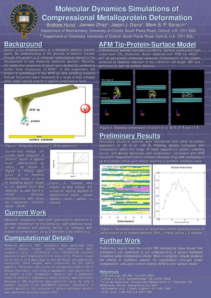
Molecular Dynamics Simulations of Compressional Metalloprotein DeformationAndrew Hung1, Jianwei Zhao2, Jason J. Davis2, Mark S. P. Sansom11Department of Biochemistry, University of Oxford, South Parks Road, Oxford, U.K. OX1 3QU2Department of Chemistry, University of Oxford, South Parks Road, Oxford, U.K. OX1 3QU Background Azurin, a Cu metalloprotein, is a biological electron transfer agent. An understanding of the process of electron transfer through this protein is of immense technological interest in the development of new molecular electronic devices1. Recently, the conduction properties of azurin were studied by conducting atomic force microscopy (C-AFM)2. In this experiment, the protein is immobilised to the AFM tip, and tunneling currents through the protein were measured at a range of bias voltages while under various extents of applied compressive force. AFM Tip-Protein-Surface Model 3-dimensional periodic boundary conditions. Surface constructed from united-atom CH4 molecules. Azurin obtained from PDB file 4AZU5 , with all non-protein molecules removed. Compression of the protein achieved by stepwise reduction of the z-direction cell length. MD runs performed at each tip-surface distance. Figure 3. Stepwise compression of azurin at (z) 42 Å, 27 Å and 17 Å Preliminary Results Secondary structural features were maintained from initial tip-surface separation of 42 Å to ~25 Å. Packing density increases with compression within this range. At lower separations, protein unfolding occurs, and density decreases with compression. Consistent with AFM conduction experiments which showed decrease in φ0 with compression up to a certain critical point before reaching a constant, minimum value. Figure 1. Schematic of a typical C-AFM experiment2. Current-bias voltage I(V) curves were acquired at different values of applied force2 (differentiated by colour), as shown in Figure 2. Fitting each curve to a modified Simmons model, a curve of tunneling barrier height φ0vs. applied force was obtained. φ0was found to initially decrease monotonously with force, but becomes constant above ~30nN. Figure 2. Current with respect to bias voltage I(V) curves for varying degrees of applied force. (black = lowest applied force, yellow = highest) Current Work Molecular simulations have been performed to determine a possible mechanism for this behaviour, with particular focus on the structure and packing density (ρ) changes with respect to compression, as φ0is believed to be related to ρ. Figure 4. Secondary structure (a) and protein atomic packing density (b) as a function of tip-surface distance. Red = α-helix, yellow = β-strands Computational Details Molecular dynamics (MD) simulations were performed under constant particle number, volume and temperature (NVT) conditions using GROMACS3 The GROMOS96 forcefield parameters were employed with time steps of 2 fs. Potential energy cut-off radii of 10 Å were used for van der Waals’ and electrostatic interactions. Bond lengths were constrained via the LINCS algorithm. The protein was coupled to a temperature bath at 300 K. Energy minimisation and 100ps of equilibration were performed on the protein at each compression distance, with a subsequent 100ps of simulation collected on which analyses were performed. Analyses of MD trajectories were performed using the suite of software included in the GROMACS software. Visualisation of system geometries and evaluation of protein secondary structure were performed using the program VMD4. Further Work Preliminary results from the current MD simulations have shown that simulations can contribute to our understanding of protein-mediated tunneling under compressive stress. Work in progress include studying the effects of hydration waters, Cu coordination changes under compression, and using a more realistic AFM tip and surface model. References 1 R. Rinaldi et al., Adv. Mat. 14, p1453 (2002) 2 J. Zhao, J. J. Davis, Nanotechnology 14(9), p1023 (2003) 3 D. van der Spoel et al., Gromacs User Manual version 3.1, Groningen, The Netherlands, Internet : www.gromacs.org (2002) 4 W. Humphrey et al., J. Molec. Graphics 14(1), p33 (1996) 5 H. Nar et al., J. Mol. Biol. 218, p427 (1991)


