What is a neutralizing antibody? (NAb)
350 likes | 568 Vues
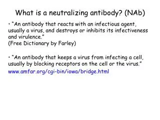
What is a neutralizing antibody? (NAb). “An antibody that reacts with an infectious agent, usually a virus, and destroys or inhibits its infectiveness and virulence.” (Free Dictionary by Farley)

What is a neutralizing antibody? (NAb)
E N D
Presentation Transcript
What is a neutralizing antibody? (NAb) “An antibody that reacts with an infectious agent, usually a virus, and destroys or inhibits its infectiveness and virulence.” (Free Dictionary by Farley) “An antibody that keeps a virus from infecting a cell, usually by blocking receptors on the cell or the virus.” www.amfar.org/cgi-bin/iowa/bridge.html
Why don’t antibodies block HIV from infecting and/or clear HIV? Free virus and infected cells both display cell-surface spikes. Why doesn’t the body make effective anti-spike antibodies to prevent infection of host cells and clear free virus and virally-infected cells?
Antibody could sterically interfere with viral attachment and/or fusion gp41 gp120 Note the trimeric spikes shown on this and subsequent slides are models, not atomic resolution structures. Envelope spike IgG CD4 CCR5 Poignard et al. Annu. Rev. Immunol., 19: 253-274, 2001. Parren and Burton Adv. Immunol.,77:195-262, 2001.
Need to understand role of gp120/gp41 in HIV entry into cells to understand why most antibodies are ineffective against HIV gp41 gp120 HIV spike (trimer of gp120/gp41 heterodimers) binds to CD4, then to CCR5 (co-receptor) on target cell. receptor co-receptor Note that CD4 acts as the receptor for HIV, but it acts as a co-receptor in T cell-mediated cellular immune responses (in which case, the T cell receptor is the receptor).
HIV Envelope glycoprotein (gp160 -->gp120/gp41) gp160 assembles as trimer, then is cleaved into gp120 and gp41 during transport to surface of infected cell gp120 (soluble, no membrane-spanning region) contains CD4 and co-receptor (CCR5 or CXCR4) binding sites gp41 is membrane-bound Cleavage of gp160 allows conformational changes upon binding CD4 and co-receptor Binding to CD4 triggers conformational changes in gp120 that open up binding site for co-receptor Co-receptor binding leads to dissociation of gp120 from membrane-anchored gp41; gp41 then refolds resulting in fusion of viral and target cell membranes
Available 3D structures Unliganded gp120 monomer (no CD4) gp120 monomer + CD4 + antibody Fab gp120 monomer + antibody Fab gp41 post-fusion state No high resolution structures of gp120 trimers or a gp120/gp41 complex
gp120* conformational states Zhou, T., et al., Nature445, 732 (2007) *Crystal structures are presently available for gp120 monomers only -- no high resolution structural information about the trimeric spike. http://www.childrenshospital.org/cfapps/research/data_admin/Site142/mainpageS142P3.html
Modeled gp120 trimer on membrane Note: HIV envelope spikes are trimers of gp120/gp41 (gp41 anchors gp120 to the viral membrane and is responsible for fusion to the host cell membrane). gp41 not shown in this model because there are no structures of gp41 in a pre-fusion state. Viral membrane Kwong et al., 2005, Science 310: 1025-1028
Trimeric HIV envelope spike structures have been examined on viruses using cryoelectron tomography The HIV envelope spike is a trimer of gp120/gp41* heterodimers. *gp (glycoprotein) 120 is 120 kDa; gp41 is 41 kDa. 3D reconstruction of SIV virions by cryo-electron tomography Zanetti, G., et al., PLoS Pathogens2, e83 (2006)
HIV-1 spike trimer structure from electron cryotomography gp41 gp41 Liu et al., 2008, Nature 455: 109-113
gp120/gp41 evade antibodies in several ways • Broadly neutralizing antibodies against gp120 or gp41 rarely found in infected patients • Sequence variability in gp120 • gp120 is shed -- shed gp120 is conformationally different than trimeric gp120 spikes on the viral membrane. Shed gp120 acts as decoy because antibodies bind more tightly to it than to envelope (membrane-bound) gp120. • gp120 carbohydrates hide protein epitopes (gp120 is 50% N-linked carbohydrate). • Antibodies are too big to access conserved regions of gp120/gp41 spikes on a virion. • CD4 binding site on gp120 is a narrow pocket. • Antibodies can’t access CCR5 binding site when HIV is bound to a target cell membrane via CD4. • gp41 is inaccessible until gp120 dissociates.
Sequence variability: A variable loop on gp120 encourages the formation of strain-specific antibodies gp120
HIV keeps the immune system busy making ineffective antibodies Infected cells release misfolded or incompletely assembled gp120s as decoys
Most anti-gp120 antibodies don’t bind to gp120 trimers on a virus NAb Antibodies produced against gp120 aren’t very useful against viruses because of “original antigenic sin”: once antibody responses are generated by vaccination (e.g., by shed gp120 or inactive gp120), host can’t make de novo Ab responses to envelope gp120 or gp120 in related viruses -- now have Abs with high affinity to shed gp120 or misfolded gp120, but low affinity to envelope gp120.
gp120 gp41 EM image of SIV (HIV shows fewer spikes) Clicker question: Env spikes are visible on free virus, but poorly accessible to antibodies, partly because gp120 is covered with carbohydrates. Carbohydrates are poorly immunogenic because: 1) They are synthesized by host machinery 2) They are charged 3) They are hydrophobic 4) They are sweet
Clicker question: Although the co-receptor binding site on gp120 (i.e., the CCR5 binding site) is highly conserved, antibodies that bind to it are generally unable to neutralize the virus. This is because… 1) Blocking the co-receptor binding site won’t prevent infection if CD4 can still bind. 2) The co-receptor binding site becomes accessible only after gp120 binds CD4 and is close to the membrane, so antibodies simply can’t fit. 3) Antibodies that recognize the co-receptor binding site are unstable and easily degraded.
Clicker question: Although the co-receptor binding site on gp120 (i.e., the CCR5 binding site) is highly conserved, antibodies that bind to it are generally unable to neutralize the virus. This is because… 1) Blocking the co-receptor binding site won’t prevent infection if CD4 can still bind. 2) The co-receptor binding site becomes accessible only after gp120 binds CD4 and is close to the membrane, so antibodies simply can’t fit. 3) Antibodies that recognize the co-receptor binding site are unstable and easily degraded. Labrijn, A. F., et al. (2003) J Virol 77, 10557-65.
In vitro neutralization assays Evaluate infectivity of HIV in the presence of different concentrations of an Ab. Which Ab is most effective at neutralization? 1) Red 2) Blue3) Green
Smaller versions of antibodies against the CCR5 binding site neutralize HIV more effectively than intact antibodies These are idealized data from an in vitro neutralization assay. scFv (single-chain Fv) Intact IgG antibody
The conserved coreceptor binding site is exposed on gp120 after CD4 binds, but there isn’t space for an intact antibody to fit both underneath gp120 and between the viral and target cell membranes One subunit of gp120 Fab Fab Fc Burton et al. (2005) PNAS 102, 14943-8
Steric restrictions on antibody access to the coreceptor binding site on gp120 scFv and Fab versions of anti-CCR5 binding site antibodies can fit in space between gp120 and target cell membrane. Intact IgG is too big. Explains why scFv and Fabs of anti-CCR5 binding site antibodies neutralize virus better than IgG versions. Burton et al. (2005) PNAS 102, 14943-8
Burton, Dennis R. et al. (2005) Proc. Natl. Acad. Sci. USA 102, 14943-14948 HIV-1 envelope spike A limited number of potent, broadly NAbs have been isolated from HIV-infected individuals Broadly NAbs (until 2009): 4E10 2F5 b12 2G12
New broadly NAbs are more potent PG9/PG16 NAbs against variable loops. Trimer-specific. Walker et al., 2009, Science 326: 289
VRC01, a new anti-CD4 binding site NAb, is more potent than b12 Wu et al., 2010, Science 329: 856
Summary of broadly NAbs available 2010 Walker & Burton, 2010
Conformational changes caused by CD4 binding to gp120 Glycosylated unliganded SIV gp120 core Deglycosylated CD4-bound HIV gp120 core Chen et al., 2005, Nature 433: 834-841
gp120 structure from Kwong et al non-neutralizing face: poorly accessible on trimer silent face: heavily glycosylated outer domain CD4 binding site neutralizing face V3 V1/V2 coreceptor binding site V2
gp120 spikes are covered in carbohydrates. Carbohydrates are poorly or nonimmunogenic. Viral membrane gp41 shown as pink cylinders Model of gp120 trimer based on structure of monomeric gp120. Carbohydrates shown in yellow or blue stick representation. Locations of neutralizing antibody epitopes indicated. Burton et al. (2005) PNAS 102, 14943-8
The resulting structure is low resolution, but shows that the trimer should be accessible to antibodies Zanetti, G., et al., PLoS Pathogens2, e83 (2006) Zhu, P. , et al., Nature441, 847 (2006)
Clicker question: Like the binding site on rhinovirus (common cold virus) for its cellular receptor, the CD4 binding site on gp120 is a narrow pocket. What is the evolutionary advantage for a virus of using a narrow pocket for a receptor binding site? Recessed pockets allow for stronger binding interactions. Cellular receptors tend to fold into pointed structures. Antibody surfaces are too broad to access them, so the virus can avoid the humoral immune response.
Clicker question: Like the binding site on rhinovirus (common cold virus) for its cellular receptor, the CD4 binding site on gp120 is a narrow pocket. What is the evolutionary advantage for a virus of using a narrow pocket for a receptor binding site? 1) Recessed pockets allow for stronger binding interactions. 2) Cellular receptors tend to fold into pointed structures . 3) Antibody surfaces are too broad to access them, so the virus can avoid the humoral immune response. Luo, et al. 1987. Science. 235: 182-91.