Download
1 / 26
260 likes | 565 Vues
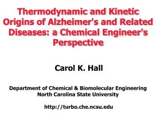
Thermodynamic and Kinetic Origins of Alzheimer's and Related Diseases: a Chemical Engineer's Perspective Carol K. Hall Department of Chemical & Biomolecular Engineering North Carolina State University http://turbo.che.ncsu.edu. Protein Folding: The ABCs.

E N D
Thermodynamic and Kinetic Origins of Alzheimer's and Related Diseases: a Chemical Engineer's PerspectiveCarol K. HallDepartment of Chemical & Biomolecular Engineering North Carolina State University http://turbo.che.ncsu.edu
Protein Folding: The ABCs A. A protein is a chain of amino acid residues arranged in a unique sequence.
C. Physiological proteins exist in the folded or “native” state, the state with the lowest free energy. Villin headpiece protein
Folded (moderate T or low denaturant) D. Proteins unfold into a “random coil” if temperature raised or denaturant (urea, GuHCl) added. E. Of all the forces thought to govern protein folding, hydrophobicity and hydrogen bonding are considered most important. Unfolded (high T or high denaturant) • www.sas.upenn.edu
Disease Pick’s Alzheimer’s Parkinson’s Prion disease (e.g. Mad Cow) Amyloid Lateral Sclerosis ( Lou Gehrig’s) Huntington’s Disease Protein tau A-beta alpha synuclein prion protein TDP-43 Huntingtin Amyloidoses:Diseases characterized by the abnormal aggregation of proteins into ordered structures, called “fibrils” or “amyloid.”
Alzheimer’s Disease • 100 years ago --Dr. Alois Alzheimer described abnormal clumps in brain of deceased dementia patient, Auguste D. • Clinical symptoms: severe dementia, loss of memory & motor skills----> death • Late onset disease : 5-10% of 65-74 year olds, 50% of 85+ year olds • 4.5 million Americans • Costs $100 billion/year • US Research Budget $650 million/year.
Protofilament structure Structure of Amyloid Fibrils Fibrils are ordered aggregates of peptides characterized by cross-beta structure AFM on fibrils of A-ß protein -sheets in a protofilament
Issues in Amyloid Disease Research • Identity of toxic species--- early oligomers or fibrils? • Kinetics of fibril nucleation and growth • Structure of fibrils • Interactions with inhibitors
Objective To develop a computational tool that : allows investigation (particularly visualization) of spontaneous fibril formation. reveals the basic physical principles underlying fibril formation . Six Blind Men and Elephant
Polyalanine– A Model System for Studying Protein Fibrillization Speculation - fibril formation is natural consequence of peptide geometry, hydrogen-bonding capability and hydrophobic interactions under slightly-denatured, concentrated conditions. Polyalanine peptides form fibrils in vitro at high concentrations (C > 1.5 mM) and high temperature (T > 40oC) (Blondelle et al., Biochem. 1997). Peptide Sequence: KA14K alpha-helix beta-sheets in a fibril
Molecular Dynamics Simulations of Protein Folding Packages: Amber, CHARMm, ENCAD, ECEPP, Discover, UNRES, etc. Force fields: describe interactions between all atoms on protein and in solvent at atomic resolution Desired Output: “folding” trajectory of a protein Limitation: very difficult to simulate folding of a single protein even with the fastest computers Implications : sacrifice details to study protein aggregation
Discontinuous Molecular Dynamics Traditional MD: Forces based on Lennard Jones (LJ) potential. Follow particle trajectories by numerically integrating Newton’s 2nd law every picosecond. Discontinuous MD: Forces field based on square-well potential. Follow particle trajectories by analytically integrating Newton’s 2nd law Particles move linearly between collisons, capture or bounce
CH3 CaH CO NH PRIME (Protein Intermediate Resolution Model): • United atom: NH, CaH, CO, R R= CH3 for alanine • Steric Interactions: hard spheres with realistic diameters • Pseudo-bonds maintain: ideal backbone bond angles trans-configuration residue L-isomerization • Covalent bond and pseudo-bond lengths set to ideal experimental values Smith and Hall. PROTEINS (2001) 44 344 Nguyen et al. Protein Sci (2004) 13 2909-2924
Model Forces: Hydrogen Bonding Hydrogen bonds between backbone amine and carbonyl groups are modeled with a directional square-well attraction of strength eH-bonding. CH3,i COi CaHi NHi COj Square-well attraction CaHj NHj Define reduced temperature as: T*=kBT/εH-bonding
Model Forces: Hydrophobic Interactions • Solvent effects captured implicitly . • Hydrophobic side chains cluster together to avoid water • Hydrophobic interaction modeled as square-well attraction between side chains. • R= εhydrophobicity/εH-bonding
Folding of Single KA14K Chain Nguyen,Marchut & Hall Biophys. J (2004)
A Constant-Temperature Simulation: 48 Peptides at c=10.0mM, T*=0.14 Nguyen & Hall, PNAS (2005)
Equilibrium Simulations: 96 Peptides Use the replica-exchange method to simulate 96-peptide systems at different temperatures and peptide concentrations. These trends qualitatively agree with experimental data (Blondelle 1997) Nguyen & Hall Biophys. J. (2004)
Fibril Structure: Intra-sheet Distance Intra-sheet distance: 4.92 ± 0.01A, comparable to experimental values of 4.76A (Shinchuk et al., Proteins, 2005)
Fibril Structure: Inter-sheet Distance Inter-sheet distance: 7.52 ± 0.23A, comparable to experimental values of 5.4A (Shinchuck et al., Proteins, 2005)
Fibril Structure: Peptide Orientation Most peptides are in-register, same as experimental results for the A-ß(10-35) peptide (Benzinger et al., PNAS 1998)
Forming Various Structures versus t* c=5mM, T*=0.14 • Amorphous aggregates form instantaneously, followed by ß-sheets, and then fibrils after a delay, called the lag time. • Appearance of a lag time indicates that this is a nucleated phenomenon. all aggregates Nguyen & Hall, J. Biol. Chem (2005)
Fibril Formation in Seeded and Unseeded Systems at T*=0.14, c=2mM Adding a seed eliminates the fibril formation lag time, as found experimentally.
In Conclusion---Technical • First intermediate resolution simulations of spontaneous “fibril” formation • Our results qualitatively agree with experimental data in general, and specifically with those obtained by Blondelle et al. (Biochemistry, 1997) on polyalanines. • Next step: Extending PRIME to all 20 amino acids. Which road to take?????
Acknowledgements • Dr. Hung D. Nguyen • Dr. Alexander J. Marchut • Dr. Anne V. Smith • Dr. Hyunbum Jang • Dr. Andrew J. Schultz • National Institutes of Health • National Science Foundation