Regulation of Cell Cycle
430 likes | 1.72k Vues
Regulation of Cell Cycle. • The entry into and the exit from the cell cycle are controlled by multiple extra-cellular signals (nutrition, mitogens , growth factors etc.)

Regulation of Cell Cycle
E N D
Presentation Transcript
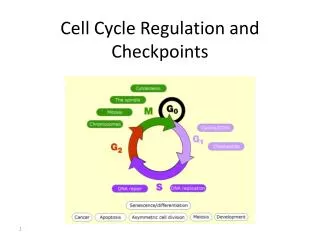
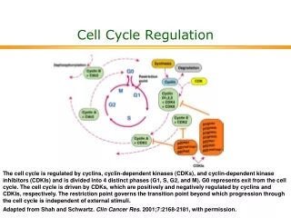
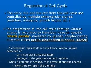
Regulation of Cell Cycle • The entry into and the exit from the cell cycle are controlled by multiple extra-cellular signals (nutrition, mitogens, growth factors etc.) • The progression of the cell cycle through various phases is regulated by transition through specific ‘check points’, mediated by specific phophorylating enzymes called cyclin-dependent kinases (CDKs) - A checkpoint represents a surveillance system, allows detection of - an incomplete previous step - damage to the genome / mitotic spindle - When a damage is sensed, cells arrest at specific phases - allow time to repair the damage
Three Major Regulatory Pathways • Start Checkpoint • The key checkpoint which determines whether or not the cell will duplicate • Regulated by growth factors, nutrients and integrity of DNA • Rb is the primary regulatory protein of this checkpoint • G2/M Checkpoint • G2 to M transition is blocked until all the DNA is duplicated • Controlled by MPF (maturation promoting factor) is blocked until DNA replicated • Spindle Checkpoint • Metaphase to anaphase transition • Makes sure that all the chromosomes are properly attached to microtubules • Anaphase-promoting complex is a key regulatory factor of this checkpoint
Growth Factors • Stimulatory growth factors • Activate the Ras pathway • Ras mutations commonly found in pancreatic, colon, lung, and bladder cancers, and occurs in about 25-30% of all cancers • Activate the PI3 kinase-Akt pathway • PTEN mutations found in 50% prostrate cancer, 35% uterine cancer and also to varying extent in other cancers • Inhibitory growth factors act through Cdk inhibitors • TGF-beta causes an increase in Cdk inhibitor p15 and p21 • The Cdk inhibitor p21 plays a key role in preventing cells containing damaged DNA from passing through the G1 checkpoint
Check-points, CDKs • Each CDK phosphorylates and thereby modulates the activity of a subset of target proteins specific for individual transition within the cell cycle • e.g. S-phase CDK may phosphorylate proteins involved in DNA replication • Mammalian cells have several CDKs, viz. • cdc2 (= CDK1), CDK2, CDK3, CDK4, CDK6, and CDK7 • act at different transitions in the cell cycle • Studies of Cdk’s and cyclins in genetically modified mice reveal a high level of plasticity, allowing different cyclins and Cdk’s to compensate for the loss of one another. • Cdk1 is capable of substituting for the all the other Cdk’s.
CDKs & Cyclins • Activation of CDKs require their association with another group of proteins called ‘Cyclins’ • Cyclin D, H, E, A, B • contribute to CDK substrate specificity • The levels of different cyclins vary during the cycle • e.g. Cyclin E accumulates in late G1, associates with CDK2, and is destroyed as cells enter S phase • The activity of the Cyclin-CDK complex is further subject to positive or negative regulation • Phosphorylation/dephosphorylation • Inhibitors of CDKs (e.g. p21 etc.) • Proteolysis of cyclins and inhibitors (APC/C)
Four Mechanisms of Cdk regulation • 1. Association of Cdk’s and cyclin partners—formation of specific Cdk/cyclin complexes is controlled by cyclin synthesis and degradation. • 2. Activation of Cdk/cyclin complexes requires phosphorylation of threonine around position 160. • This is catalyzed by an enzyme called CAK (for Cdk-activating kinase), which is composed of Cdk7/cyclin H. • 3. Inhibitory phosphorylation of tyrosine near the Cdk amino terminus, catalyzed by the Wee1 protein kinase. • The Cdk’s are then activated by dephosphorylation by Cdc25 protein phosphatases. • 4. Binding of inhibitory proteins Cdk inhibitors (CKIs). • In mammalian cells, there are two families of Cdk inhibitors: Ink4 and Cip/Kip
The regulation of Cdk activity by inhibitory phosphorylation • The active cyclin-Cdk complex is turned off when the kinase Wee 1 phosphorylates two closely spaced sites above the active site. • Removal of these phosphates by the phosphatase Cdc25 activates the cyclin-Cdkcomplex
CDK inhibitor proteins (CDKI) • The cyclin D/CDK4/CDK6, Cyclin E/CDK2, and Cyclin A/CDk2 are inhibited by a group of CDK inhibitor proteins (CDKI) that include p21 & p27. • CDKIs also impair CDK activating kinase activity (CAK)
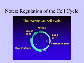
Restriction point control : G1 to S progression • The transition from G1 to S phase is regulated by a checkpoint called “Restriction point” or Start - a very important regulatory step, to ensure repair of genome damage before initiation of DNA replication • The initiation of cycle, i.e. entry of cells into G1 phase, is determined by extra-cellular signals (mitogens, nutrients & growth factors) • Growth factors induce synthesis of D-type cyclins (D1, D2, D3) • D cyclins associate with CDK4 and CDK6 in G1 • Cyclin D/CDK4/CDK6 complexes are activated through phosphorylation by an enzyme complex called CAK (CDK activating kinase)
Induction of D-type cyclins • Proliferation of animal cells is regulated largely by extracellular growth factors that control progression through the restriction point in late G1. • The activity of cyclin-CDK complexes is tightly regulated by CDK inhibitors. Some Growth factors shut off production of these inhibitors. • Growth factors stimulate cyclin D1 synthesis through the Ras/Raf/MEK/ERK pathway, and are synthesized as long as growth factors are present. • Cyclin D1 is also rapidly degraded, so the intracellular concentration rapidly falls if growth factors are removed.
Cyclin D & Cell Cycle Progression As long as growth factors are present through G1, Cdk4,6/cyclin D1 complexes drive cells through the restriction point. Defects (overexpression) in cyclin D1 regulation could contribute to the loss of growth regulation characteristic of cancer cells. Many human cancers arise as a result of defects in cell cycle regulation.
Rb controls the G1 to S transition • The Retinoblastoma protein, Rb, is phosphorylated by Cyclin D/CDK4/CDK6, which is necessary to drive the cell past the restriction point • Once the cell crosses the restriction point, mitogenic stimulation is no longer needed, and the entry into the S phase is ensured • In its hypophosphorylated state, the Rb is complexed with E2F family of transcription factors (E2F1 – E2F5) • Phosphorylation of Rb dissociates E2F, which then heterodimerizes with DP family of transcription factors (DP-1, DP-2, DP-3)
Cell cycle regulation of Rb and E2F • Rb plays a key role in coupling cell cycle machinery to the expression of genes required for cell cycle progression. • In G0 or early G1, Rb binds to E2F transcription factors, which suppresses expression of genes involved in cell cycle progression. • Rb is phosphorylated by Cdk4,6/cyclin D complexes as cells pass through the restriction point, and dissociates from E2F, allowing transcription to proceed.
Restriction point control and S phase • Cyclin E/CDK2 kinase is needed to maintain Rb in its hyperphosphorylated state • As Cyclin E/CDK2 activity decreases, cyclin A synthesis is induced. • Accumulation of the Cyclin A/CDK2 complexes signals entry into S phase.
G2 to M Checkpoint • The mitotic Cdk1-cyclin B complex (MPF) controls the G2 checkpoint by phosphorylating proteins involved in the early stages of mitosis • MPF = Maturation-promoting factor • Activated by multi-step process • MPF phosphorylateslamin proteins of the nuclear lamina (causing breakup of nuclear membrane) • MPF phosphorylatescondensin complex which may trigger chromosome condensation
MPF regulation • MPF is regulated by phosphorylation and dephosphorylation of Cdk1. • Cyclin B is synthesized and forms complexes with Cdk1 during G2. • Cdk1 is phosphorylated and inhibited, leading to accumulation of inactive Cdk1/cyclin B complexes throughout G2. • Dephosphorylation activates Cdk1, which phosphorylates several proteins that initiate the events of M phase. • Cyclin B is degraded by ubiquitin-mediated proteolysis.
DNA damage checkpoints • DNA damage checkpoints ensure that damaged DNA is not replicated and passed on to daughter cells. • The cell cycle is arrested in response to damaged or unreplicated DNA.
Arrest at the DNA damage checkpoints • DNA damage checkpoints are mediated by related protein kinases, ATM and ATR, that are activated in response to DNA damage. • They activate a signaling pathway that leads to cell cycle arrest, DNA repair, and sometimes, programmed cell death. • ATM is activated by double-strand breaks, ATR is activated by single-stranded or unreplicated DNA. • They phosphorylate and activate the checkpoint kinasesChk2 and Chk1. • Chk1 and Chk2 phosphorylate and inhibit Cdc25 phosphatases, which are required to activate Cdk1 and Cdk2. • Inhibition of Cdk2 results in cell cycle arrest in G1 and S. • Inhibition of Cdk1 results in arrest in G2.
Mdm2 controls the levels of p53 in the nucleus • Mdm 2 controls the level of p53 in the nucleus by binding p53 and shuttling it out of the nucleus where it is destroyed by the ubiquitin-dependent pathway • If p53 is phosphorylated by the ATM/ATR pathway and is acetylated, it will no longer interact with Mdm2 • Thus, the levels of p53 increase in the nucleus • P53 levels also increase if there is excessive stimulation of mitogenic pathways • Mitogen activated pathways lead to up-regulation of transcription factor Myc • Myc causes Arf to be produced • Arf inactivates Mdm2 • No Mdm2 results in increased p53 levels
***p53 *** • p53 plays a pivotal role in cell cycle arrest in response to DNA damage • When DNA is damage ATM & ATR (two DNA-dependent protein kinases) leads to the phosphorylation of p53, preventing its degradation • Thus, DNA strand breaks by UV or ionizing radiation increase levels of p53 protein • P53 is a transcription factor • p53 then stimulates the expression of p21 (a CDK inhibitor) that causes cell cycle arrest!!!!! • p21 blocks cdk/cyclin complexes
Role of p53 in G1 arrest • In mammalian cells, arrest at the G1 checkpoint is also mediated by protein p53, which is phosphorylated by both ATM and Chk2. • p53 is a transcription factor, and its increased expression leads to induction of Cdk inhibitor p21. • p21 inhibits Cdk2/cyclin E complexes, leading to cell cycle arrest in G1. • The gene encoding p53 is frequently mutated in human cancers. • Loss of p53 prevents G1 arrest in response to DNA damage, so the damaged DNA is replicated and passed on to daughter cells.
Li-Fraumeni Syndrome (LFS) • Rare “Cancer Families” with a history of many different forms of cancer (bone and soft tissue sarcoma, breast cancer, brain tumor, leukemia, and adrenocortical carcinoma) • Usually afflict patient at an unusually early age • Inherited in an autosomal dominant pattern • > 70% of families have a mutant form of the TP53 gene (codes for p53) • LFS can occur both as a sporadic and familial form • Mutations in both alleles are necessary to inactivate the TP53 gene