
Vinculin
Borisova et al., Fig. S1. Vinculin WT. Vinculin -/-. Vinculin. active 1 integrin. overlay.

Vinculin
E N D
Presentation Transcript
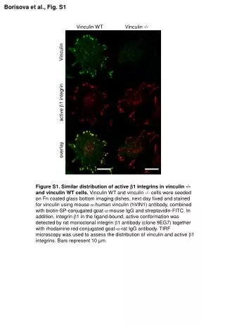
Borisova et al., Fig. S1 Vinculin WT Vinculin -/- Vinculin active 1 integrin overlay Figure S1. Similar distribution of active 1 integrins in vinculin -/- and vinculin WT cells. Vinculin WT and vinculin -/- cells were seeded on Fn coated glass bottom imaging dishes, next day fixed and stained for vinculin using mouse -human vinculin (hVIN1) antibody, combined with biotin-SP-conjugated goat -mouse IgG and streptavidin-FITC. In addition, integrin 1 in the ligand-bound, active conformation was detected by rat monoclonal integrin 1 antibody (clone 9EG7) together with rhodamine red conjugated goat--rat IgG antibody. TIRF microscopy was used to assess the distribution of vinculin and active 1 integrins. Bars represent 10 µm.