Download

1 / 32
370 likes | 963 Vues
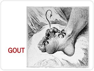
Diagnosis of gout. Background. Gout is a severe inflammatory disease caused by the deposition of monosodium urate (MSU) crystals in joints and other tissues Gout is the most frequent inflammatory arthritis in men The i ncidence and prevalence of gout are rising in post-menopausal women

E N D
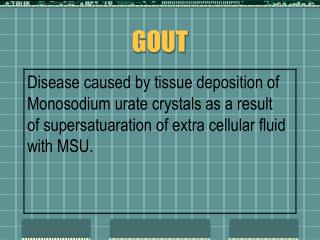
Background • Gout is a severe inflammatory disease caused by the deposition of monosodium urate (MSU) crystals in joints and other tissues • Gout is the most frequent inflammatory arthritis in men • The incidence and prevalence of gout are rising in post-menopausal women • 1-2% of adults are affected • The prevalence of gout increases with age • Gout is often misdiagnosed or diagnosed late in its clinical course Lawrence RC, et al. Arthritis Rheum 1998;41:778-799. Mikuls TR, et al. Ann Rheum Dis 2005;64:267-272. Zhang W et al, Ann Rheum Dis, 2006;65:1301-1311
EULAR evidence based recommendations for gout Zhang W, et al. Ann Rheum Dis 2006;65(10):1301-1311.
1 2 EULAR recommendations 2006 for gout: diagnosis In acute attacks the rapid development of severe pain, swelling and tenderness that reaches its maximum within just 6-12 hours, especially with overlying erythema, is highly diagnostic of crystal inflammation though not specific for gout. For typical presentations of gout (such as recurrent podagra with hyperuricemia) a clinical diagnosis alone is reasonably accurate but not definitive without crystal confirmation. Zhang W, et al. Ann Rheum Dis 2006;65(10):1301-1311.
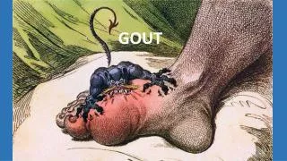
Acute gout: classical clinical picture • Acute, very painful,monoarticular inflammation usually affecting the big toe (podagra)(70%) (less frequently other foot joints, ankle, knee, finger, wrist, elbow) • Typical rapid development of severe pain, swelling and tenderness that reaches its maximum within just 6-12 hours, especially with overlying erythema • As inflammation disappears, the skin over the joint often peels • Attacks often start at night or in the early morning • Attack usually resolve within 5-10 days By kind permission of L. Punzi, Rheumatology Unit, University of Padua Wallace SL, et al. Arthritis and Rheumatism 1977;20(3):895-900.
Often typical gout is gout – but not always • Not rarely what does not look like gout is gout Differential diagnosis of big toe gout (podagra) • Pseudogout (acute calcium pyrophosphate arthropathy) • Pseudogout-like (basic calcium phosphate) (pseudopodagra) • Reactive arthritis • Psoriatic arthritis • Septic arthritis • Sarcoidosis • Others Zhang W, et al. Ann Rheum Dis 2006;65:1301-11. Richette P, et al. Lancet 2010;375:318-328.
Differential diagnosis of ankle gout • Sarcoidosis • Reactive arthritis • Psoriatic arthritis • Enteroarthritis • Septic arthritis • Other crystal-induced arthritides • acute pyrophosphate arthropathy (pseudogout) • basic calcium phosphate (pseudogout-like) By kind permission of L. Punzi, Rheumatology Unit, University of Padua Zhang W, et al; Ann Rheum Dis 2006;65:1301-1311. Richette P, et al. Lancet 2010;375:318-328. De Leonardis F, et al. Rheumatol Int 2007;28:1-7.
Differential diagnosis of elbow gout or bursitis • Septic arthritis or bursitis • Post-traumatic bursitis • Rheumatoid nodules with bursitis • Other crystal-induced arthritides or bursitis • acute pyrophosphate arthropathy (pseudogout) • basic calcium phosphate (pseudogout-like) By kind permission of L. Punzi, Rheumatology Unit, University of Padua Ning TC, et al. Curr Opin Rheumatol 2010;22(2):181-187. .
Differential diagnosis of hand-wrist gout • Algoneurodystrophy • Pitting oedema • polymyalgia rheumatica • psoriatic arthritis • Septic arthritis • Other crystal-induced arthritides or bursitis • acute pyrophosphate arthropathy (pseudogout) By kind permission of L. Punzi, Rheumatology Unit, University of Padua Richette P, et al. Lancet 2010;375:318-328. De Leonardis F, et al. Rheumatol Int 2007;28:1-7. Ning TC, et al. Curr Opin Rheumatol 2010;28:181-187.
Chronic arthritis: gout or not gout? Gout or psoriatic arthritis? Gout or rheumatoid arthritis? Gout or osteoarthritis (Heberden’s nodes)? By kind permission of L. Punzi, Rheumatology Unit, University of Padua
3 4 EULAR recommendations 2006 for gout: diagnosis Demonstration of monosodium urate (MSU) crystals in synovial fluid or tophus aspirates permits a definitive diagnosis of gout. A routine search for MSU crystals is recommended in all synovial fluid samples obtained from undiagnosed inflamed joints. Zhang W, et al. Ann Rheum Dis 2006;65(10):1301-1311.
Likelihood ratio (HR) for various features in the diagnosis of gout Zhang W, et al. Ann Rheum Dis 2006;65:1301-1311.
Ultrasound-guided arthrocentesis Grassi W, et al. Ann Rheum Dis 1999;58:595-597.
MSU crystals are always found in: • Synovial fluid samples from inflamed joints • Previously inflamed joints of patients untreated with urate-lowering drugs • Material from tophi • Joints of treated patients… before they dissolve By kind permission of L. Punzi, Rheumatology Unit, University of Padua Zhang W, et al. Ann Rheum Dis 2006;65(10):1301-1311.
Synovial fluid analysis for crystals Steps to follow: Steps to follow: • To detect whether there are any crystals The technique for detection of MSU and CPPD differs • If so: to identify what type they are Zhang W, et al. Ann Rheum Dis 2006;65(10):1301-1311.
Monosodium Urate (MSU) • Acicular • Intense birefringence • Negative elongation axis By kind permission of L. Punzi, Rheumatology Unit, University of Padua First order red compensator Polarised light Ordinary light Identification of MSU crystals Richette P, et al. Lancet 2010;375:318-328. Pascual E, et al. Ann Rheum Dis 2009; 68:3-7. Sivera F, et al. Ann Rheum Dis 2008;67:273-275.
By kind permission of L. Punzi, Rheumatology Unit, University of Padua Polarised light First order red compensator MSU crystals from tophi Richette P, et al. Lancet 2010;375:318-328. Pascual E , et al. Ann Rheum Dis 2009;68:3-7. Sivera F, et al. Ann Rheum Dis 2008;67:273-275.
5 EULAR recommendations 2006 for gout: diagnosis Identification of MSU crystals from asymptomatic joints may allow definite diagnosis in intercritical periods. Zhang W, et al. Ann Rheum Dis 2006;65(10):1301-1311.
MSU crystals in synovial fluid during intercritical periods • Aspiration of 101 asymptomatic gouty joints • 80 knees • 21 first MTP joints • All had previously been inflamed and had been free of inflammation • over two months. • MSU crystals were found in: • 43/43joints from untreated patients • 34/48 (71%)joints from patients on urate-lowering drugs (p<0.001). Pascual E, et al. Ann Intern Med 1999;131:756-759.
6 EULAR recommendations 2006 for gout: diagnosis Gout and sepsis may coexist, so when septic arthritis is suspected gram stain and culture of synovial fluid should still be performed even if monosodium urate crystals are identified. Zhang W, et al. Ann Rheum Dis 2006;65(10):1301-1311.
7 EULAR recommendations 2006 for gout: diagnosis While being the most important risk factor for gout, serum uric acid levels do not confirm or exclude gout since many people with hyperuricaemia do not develop gout, and during acute attacks serum levels may be normal Zhang W, et al. Ann Rheum Dis 2006;65(10):1301-1311.
Normative Aging Study Follow-up of 2046 men, free of gout at the onset, over 14.9 years Campion EW. Am J Med 1987;82:421-426.
Inflammation… effects on serum UA Inflammation… effects on serum UA • During a gout attack, serum uric acid drops because of an increase of its renal excretion • For example: 50% of gouty patients have normal or low UA levels during attacks • Diseases with persistent inflammation have a negative association with hyperuricaemia and gout • For example: Rheumatoid arthritis • Serum acid uric is an inverse acute phase substance? Urano W, et al. J Rheumatol 2002;29(9):1950-1953. Agudelo CA, et al. Arthritis Rheum 1984;27(4):443-448 Wu VC, et al. Am J Kidney Dis 2005;45(1):88-95.
8 EULAR recommendations 2006 for gout: diagnosis Urinary uric acid excretion should be determined in selected gout patients, especially those with a family history of young onset gout, onset of gout under age 25, or with renal calculi Zhang W, et al. Ann Rheum Dis 2006;65(10):1301-1311.
9 EULAR recommendations 2006 for gout: diagnosis Although radiographs may be useful for differential diagnosis and may show typical features in chronic gout, they are not useful in confirming the diagnosis of early or acute gout. Zhang W, et al. Ann Rheum Dis 2006;65(10):1301-1311.
Likelihood ratio (HR) for radiographic features in the diagnosis of gout Zhang W, et al. Ann Rheum Dis 2006;65:1301-1311.
DIP PIP Big toes Radiographic features of gout By kind permission of L. Punzi, Rheumatology Unit, University of Padua
EULAR recommendations 2006 for gout: diagnosis 10 Risk factors and associated co-morbidity can be assessed during the diagnosis of gout, including features of the metabolic syndrome (obesity, hyperglycaemia, hyperlipidaemia, hypertension). Zhang W, et al. Ann Rheum Dis 2006;65(10):1301-1311.