
RAR α
A. B. PaTu-T. PaTu-S. RAR α. no Ab. Input. DR5-Typ10. HoxB4-Ex2. HoxB4-Ex1. miR-10a.

RAR α
E N D
Presentation Transcript
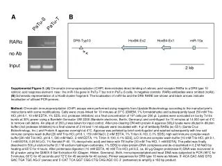
A B PaTu-T PaTu-S RARα no Ab Input DR5-Typ10 HoxB4-Ex2 HoxB4-Ex1 miR-10a Supplemental Figure 3: (A) Chromatin immunoprecipitation (CHIP) demonstrates direct binding of retinoic acid receptor RARα to a DR5 type 10 retinoic acid response element near the miR-10a gene in PaTu-T but not in PaTu-S cells. In negative controls RARα antibodies were omitted (noAb). (B) Schematic representation of a HoxB cluster fragment. The localisation of miR10a and the DR5 RARE are shown and black arrows indicate localisation of utilised PCR-primers. Method: Chromatin immunoprecipitation (ChIP) assays were performed using reagents from Upstate Biotechnology according to the manufacturer’s instructions with some modifications. Cells were cross-linked for 10 minutes at 37°C (DMEM, 1% formaldehyde) and subsequently lysed (50 mM Tris-HCl, pH 8.1, 10 mM EDTA, 1% SDS, incl. protease inhibitors) at a final concentration of 106 cells per 200 µl. Lysates were sonicated on ice by 7x10s bursts at 30% power using a Bandelin Sonicator UW 2200 (Bandelin electronic, Berlin, Germany) and centrifuged for 10 minutes at 14.000 rpm at 4°C to remove cell debris. An aliquot of 200 µl was taken for input control. After pre-clearing ON with protein A agarose 300µl lysate were diluted in dilution buffer (incl protease inhibitors) to a final volume of 2 ml and 1 ml aliquots were incubated with 4 µl of antibody RARα (sc-551) (Santa Cruz Biotechnology, Inc.) and Protein A agarose overnight at 4°C. Agarose was pelleted by brief centrifugation and washed subsequently with low salt immune complex wash buffer [20 mM Tris-HCl, pH 8.1, 150 mM NaCl, 2 mM EDTA, 1% Triton X-100, 0.1% SDS], high salt immune complex wash buffer [20 mM Tris-HCl, pH 8.1, 500 mM NaCl, 2 mM EDTA, 1% Triton X-100, 0.1% SDS], LiCl immune complex wash buffer [10 mM Tris-HCl, pH 8.1, 1 mM EDTA, 0.25 M LiCl, 1% Nonidet P-40, 1% deoxycholic acid] and twice with TE buffer [10 mM Tris-HCl, 1 mM EDTA]. The pellet was finally dissolved in 500 µl elution buffer [0.1 M sodium-hydrogen-carbonate, 1% SDS] to elute protein-DNA complexes and de-crosslinked in 0.2 M NaCl by heating at 65°C for 4 hours. After proteinase digestion (10 mM EDTA, 40 mM Tris-HCl, pH 6.5, ca. 60 µg Qiagen proteinase K) DNA was recovered in 60 µl water using the QIAEX-II Gel Extraction Kit (Qiagen, Hilden, Germany). Both, immunoprecipitated and input DNA was subjected to PCR (95°C for 5 minutes, 66°C for 45 seconds and 72°C for 45 seconds for 40 cycles). Primer sequences for DR5 type 10 were as follows: 5’-ACA GAC AAG GTG GAC TGA TGC AG-3’ (sense) and 5’-CAT TCA GGT CGG CTG CAG AGC CC-3’ (antisense) to amplify a 192 bp product. 2 kb