Download
1 / 66
700 likes | 1.02k Vues
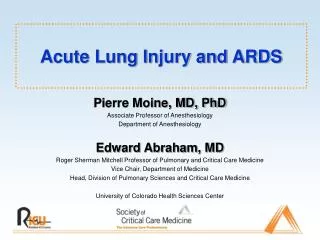
Acute Lung Injury and ARDS. Pierre Moine, MD, PhD Associate Professor of Anesthesiology Department of Anesthesiology Edward Abraham, MD Roger Sherman Mitchell Professor of Pulmonary and Critical Care Medicine Vice Chair, Department of Medicine

E N D
Acute Lung Injury and ARDS Pierre Moine, MD, PhD Associate Professor of Anesthesiology Department of Anesthesiology Edward Abraham, MD Roger Sherman Mitchell Professor of Pulmonary and Critical Care Medicine Vice Chair, Department of Medicine Head, Division of Pulmonary Sciences and Critical Care Medicine University of Colorado Health Sciences Center
Definitions • The 1994 North American-European Consensus Conference (NAECC) criteria: • Onset - Acute and persistent • Radiographic criteria - Bilateral pulmonary infiltrates consistent with the presence of edema • Oxygenation criteria - Impaired oxygenation regardless of the PEEP concentration, with a Pao2/Fio2 ratio 300 torr (40 kPa) for ALI and 200 torr (27 kPa) for ARDS • Exclusion criteria - Clinical evidence of left atrial hypertension or a pulmonary-artery catheter occlusion pressure of 18 mm Hg. Bernard GR et al., Am J Respir Crit Care Med 1994
The 1994 NAECC Definition Limitations • Descriptive definition - Permits inclusion of a multiplicity of clinical entities ranging from autoimmune disorders to direct and indirect pulmonary injury • Does not address the cause of lung injury • Does not provide guidelines on how to define acute • The radiological criteria are not sufficiently specific • Does not account for the level of PEEP used, which affects the Pao2/Fio2 ratio • Does not specify the presence of nonpulmonary organ system dysfunction at the time of diagnosis • Does not include the different specific mechanistic pathways involved in producing lung injury Atabai K and Matthay MA, Thorax 2000 Abraham E et al., Crit Care Med 2000
The 1998 NAECC Updated Recommendations • The collection of epidemiologic data should be based on the 1994 NAECC definitions. 2. The severity of ALI/ARDS should be assessed by the Lung Injury Score (LIS) or by the APACHE III or SAPS II scoring systems. 3. The factors that affect prognosis should be taken into account. The most important of these are incorporated into the GOCA stratification system. 4. It will be also useful to record: • Information relating to etiology (at a minimum, direct or indirect cause) • Mortality, including cause of death, and whether death was associated with withdrawal of care • Presence of failure of other organs and other time-dependent covariates • Follow-up information, including recovery of lung function and quality of life Artigas A et al., Am J Respir Crit Care Med 1998
Stratification System of Acute Lung Injury GOCA Artigas A, et al. Am J Respir Crit Care Med. 1998.
Epidemiology • NIH, 1972 - Incidence of ARDS in the United States: 75 cases per 105 person.years population (approximately 150,000 cases per year) • International multi-center ALI/ARDS cohort studies, 1989 - 2002 • Incidence estimates of ALI/ARDS = 1.3 to 22 cases per 105 person.years • ARDS Network Study (NAECC definitions), 2003 - Incidence of ALI/ARDS in the United States: 32 cases per 105 person.years (range 16 - 64) • ARDS Network Study (NAECC definitions), 2003 - The average number of cases of ALI per ICU bed per year (2.2) varied significantly from site to site (range 0.7 - 5.8) Goss CH et al., ARDS Network, Crit Care Med 2003
Clinical Disorders Associated with the Development of ALI/ARDS • Direct insult • Common • Aspiration pneumonia • Pneumonia • Less common • Inhalation injury • Pulmonary contusions • Fat emboli • Near drowning • Reperfusion injury • Indirect insult • Common • Sepsis • Severe trauma • Shock • Less common • Acute pancreatitis • Cardiopulmonary bypass • Transfusion-related TRALI • Disseminated intravascular • coagulation • Burns • Head injury • Drug overdose Atabai K, Matthay MA. Thorax. 2000. Frutos-Vivar F, et al. Curr Opin Crit Care. 2004.
Clinical Risk Factors Predictive of a Poor Outcome Independent predictors repeatedly associated with higher mortality rates • Severity of the illness (SAPS II and APACHE) • Non-pulmonary organ dysfunction • Comorbid diseases • Sepsis • Liver dysfunction/cirrhosis • Advanced age Other independent predictors • Late ARDS ( 48 hours after MV initiation) or length of MV prior to ARDS • Organ transplantation • HIV infection • Immunosuppression • Active malignancy • Oxygenation index (mean airway pressure x Fio2 x 100/Pao2) • Mechanisms of lung injury • Barotrauma • Right ventricular dysfunction • Fio2 (High Fio2) • Pao2/Fio2<100 mmHg/Pao2/Fio2 on day 3 • Dead-space fraction • Lower levels of PEEP or no PEEP • Late respiratory acidosis • McCabe score • Chronic alcoholism Atabai K, Matthay MA. Thorax. 2000. Ware LB. Crit Care Med. 2005. Ferguson ND, et al. Crit Care Med. 2005.
Plasma Biologic Markers Predictive of a Poor Outcome Acute inflammation Interleukin(IL)-6, IL-8 Endothelial injury von Willebrand factor antigen Epithelial type II cell molecules Surfactant protein-D Adhesion molecule Intercellular adhesion molecule-1 (ICAM-1) Neutrophil-endothelial interaction Soluble tumor necrosis factor receptors I and II (sTNFRI/II) Procoagulant activity Protein C Fibrinolytic activity Plasminogen activator inhibitor-1 Ware LB. Crit Care Med. 2005.
Mortality from ARDS • ARDS mortality rates - 31% to 74% • The variability in the rates quoted is related to differences in the populations studied and in the precise definitions used. • The main causes of death are nonrespiratory causes (i.e., die with, rather than of, ARDS). • Respiratory failure has been reported as the cause of death in 9% to 16% of patients with ARDS. • Early deaths (within 72 hours) are caused by the underlying illness or injury, whereas late deaths are caused by sepsis or multiorgan dysfunction. • There is a controversy about the role of hypoxemia as a prognostic factor in adults. Nevertheless, in some studies, both Pao2/Fio2 ratio and Fio2 were variables independently associated to mortality. Frutos-Vivar F, et al. Curr Opin Crit Care. 2004. Vincent JL, et al. Crit Care Med. 2003. Ware LB. Crit Care Med. 2005.
One-year Outcomes in Survivors of the Acute Respiratory Distress Syndrome • Persistent functional limitation • Extrapulmonary diseases (primarily):Muscle wasting and weakness (corticosteroid-induced and critical-illness-associated myopathy) Entrapment neuropathy Heterotopic ossification • Intrinsic pulmonary morbidity (5%): Bronchiolitis obliterans organizing pneumonia Bronchiolitis obliterans Herridge MS, et al. N Engl J Med. 2003.
Positive-pressure Mechanical Ventilation Currently, the only therapy that has been proven to be effective at reducing mortality in ALI/ARDS in a large, randomized, multi-center, controlled trial is a protective ventilatory strategy. Tidal volume and plateau pressure
Ventilator-induced Lung InjuryConceptual Framework • Lung injury from: • Overdistension/shear - > physical injury • Mechanotransduction - > “biotrauma” • Repetitive opening/closing • Shear at open/collapsed lung interface • Systemic inflammation and death from: • Systemic release of cytokines, endotoxin, bacteria, proteases “volutrauma” “atelectrauma”
Ventilator-induced Lung Injury Three different pathologic entities: • High-permeability type pulmonary edema • Mechanical over-inflation/distortion of lung structures • Lung inflammation “Biotrauma” Rouby JJ, et al. Anesthesiology. 2004.
Ventilator-induced Lung Injury High-permeability type pulmonary edema • Main causative factor: End-inspiratory lung volume >> peak inspiratory pressure “volutrauma more appropriate than barotrauma” • Mechanisms altering the alveolar-capillary barrier permeability during MV involve: - Increased transmural vascular pressure - Surfactant inactivation - Mechanical distortion and disruption of endothelial cells - Regional activation of inflammatory cells Rouby JJ, et al. Anesthesiology. 2004. Ricard JD, et al. Eur Respir J. 2003.
Ventilator-induced Lung Injury Mechanical overinflation/distortion of lung structures • Emphysema-like lesions, lung cysts, and bronchiectasis • These lesions predominate in nondependent and caudal lung regions • The degree of overinflation is dependent on: - Tidal volume - Peak airway pressure - Duration of mechanical ventilation - Time exposed to an Fio2 > 0.6 Rouby JJ, et al. Anesthesiology. 2004.
Ventilator-induced Lung Injury Lung inflammation “biotrauma” • Lung overinflation or overstretching produces regional and systemic inflammatoryresponse that may generate or amplify multiple-system organ failure. • Factors converting the shear stress applied to an injured lung into regional and systemic inflammation are still incompletely elucidated but could include: - Repetitive opening and collapse of atelectatic lung units - Surfactant alterations - Loss of alveolo-capillary barrier function - Bacterial translocation - Overinflation of health lung regions Rouby JJ, et al. Anesthesiology. 2004. Dreyfuss D, et al. Am J Respir Crit Care Med. 2003.
Ventilator-induced Lung Injury • Two primary mechanistic factors: • Overdistension of the alveoli by high transpulmonary pressures: volutrauma • Shear-stress forces produced by repetitive alveolar recruitment and derecruitment (collapse) • Animal data so compelling that in early 1990s the SCCM and ACCP recommended reduction in tidal volume and limiting end-expiratory plateau pressure to < 35 cm H20
Traditional Approach High priority to traditional goals of acid-base balance and patient comfort Lower priority to lung protection Low Stretch Approach High priority to lung protection Lower priority to traditional goals of acid-base balance and comfort Tidal Volume Strategies in ARDS
ARDS Net Study 01: Hypothesis In patients with ALI/ARDS, ventilation with reduced tidal volume will limit “volutrauma” and improve survival. “Lung-protective strategies” ARDS Network. N Engl J Med. 2000.
ARDS Network Low VT Trial • Patients with ALI/ARDS (NAECC definitions)of < 36 hours • Ventilator procedures • Volume-assist-control mode • RCT of 6 vs. 12 ml/kg of predicted body weight PBW Tidal Volume (PBW/Measured body weight = 0.83) • Plateau pressure 30 vs. 50 cmH2O • Ventilator rate setting 6-35 (breaths/min) to achieve a pH goal of 7.3 to 7.45 • I/E ratio:1.1 to 1.3 • Oxygenation goal: PaO2 55 - 80 mmHg/SpO2 88 - 95% • Allowable combination of FiO2 and PEEP: FiO2 0.3 0.4 0.4 0.5 0.5 0.6 0.7 0.7 0.7 0.8 0.9 0.9 0.9 1.0 1.0 1.0 1.0 PEEP 5 5 8 8 10 10 10 12 14 14 14 16 18 18 20 22 24 • The trial was stopped early after the fourth interim analysis (n = 861 for efficacy; p = 0.005 for the difference in mortality between groups) ARDS Network. N Engl J Med. 2000.
1.0 0.9 0.8 0.7 0.6 0.5 0.4 0.3 0.2 0.1 0.0 0 20 40 60 80 100 120 140 160 180 ARDS Network: Improved Survival with Low VT Proportion of Patients Lower tidal volumes Survival Discharge Traditional tidal values Survival Discharge Days after Randomization ARDS Network. N Engl J Med. 2000.
ARDS Network: Main Outcome Variables ARDS Network. N Engl J Med. 2000.
Median Organ Failure Free Days = 6 ml/kg = 12 ml/kg * * * *
ARDS Network: Additional Findings In ALI and ARDS patients, 6 ml/kg PBW tidal volume ventilation strategy was associated with: • PaO2/FiO2 lower in 6 ml/kg low VT group • High RR prevented hypercapnia with minimal auto-PEEP (difference of median intrinsic PEEP between the groups was < 1 cm H2O) • No difference in their supportive care requirements (vasopressors-IV fluids-fluid balance-diuretics-sedation) • ~10% mortality reduction • Less organ failures • Lower blood IL-6 and IL-8 levels ARDS Network. N Engl J Med. 2000. Parsons PE, et al. Crit Care Med. 2005. Hough CL, et al. Crit Care Med. 2005. Cheng IW, et al. Crit Care Med. 2005.
Ventilator-induced Lung Injury • Two primary mechanistic factors: • Overdistension of the alveoli by high transpulmonary pressures • Shear-stress forces produced by repetitive alveolar recruitment and derecruitment (collapse) - Atelectrauma • In animal models, the repetitive cycle of alveolar collapse and re-recruitment has been associated with worsening lung injury. The extent of this injury has been reduced in animals through the use of PEEP levels that prevent derecruitment at end-expiration.
VT ~ 6 ml/kg PEEP ~13-16 VT~12 ml/kg PEEP ~9 Significant prognostic factors responsible of the ventilatory treatment effect: • APACHE II score • Mean PEEP during the first 36 hours (with a protective effect) • Driving pressures (PPLAT - PEEP) during the first 36 hours Amato M, et al. N Engl J Med. 1998.
PEEP in ARDSHow much is enough ? • PEEP by avoiding repetitive opening and collapse of atelectatic lung units, could be protective against VILI • High PEEP should make the mechanical ventilation less dangerous than low PEEP. • The recruitment is obtained essentially at end-inspiration, and the lung is kept open by using PEEP to avoid end-expiratory collapse. • PEEP, by preserving inspiratory recruitment and reestablishingend-expiratory lung volume, has been shown to prevent surfactantloss in the airways and avoid surface film collapse. Levy MM. N Engl J Med. 2004. Rouby JJ, et al. Am J Respir Crit Care Med. 2002. Gattinoni L, et al. Curr Opin Crit Care. 2005.
PEEP in ARDSHow much is enough ? • “Optimal PEEP”: Allowing for a given ARDS an optimization of arterial oxygenation without introducing a risk of oxygen toxicity and VILI, while having the least detrimental effect on hemodynamics, oxygen delivery, and airway pressures. • There has never been a consensus regarding the optimum level of PEEP for a given patient with ARDS. • The potential for recruitment may largely vary among the ALI/ARDS population. • PEEP may increase PaO2 without any lung recruitment because of a decrease in and/or a different distribution of pulmonary perfusion. Levy MM. N Engl J Med. 2004. Rouby JJ, et al. Am J Respir Crit Care Med. 2002. Gattinoni L, et al. Curr Opin Crit Care. 2005.
NIH-NHLBI ARDS Network: Hypothesis In patients with ALI/ARDS (NAECC definitions)of < 36 hours who receive mechanical ventilation with a VT of 6 ml/kg of PBW, higher PEEP may improve clinical outcomes. NHLBI ARDS Clinical Trials Network. N Engl J Med. 2004.
NIH-NHLBI ARDS Network • Patients with ALI/ARDS (NAECC definitions)of < 36 hours • Ventilator procedures • Volume-assist-control mode • Tidal-volume goal: 6 ml/kg of predicted body weight PBW • Plateau pressure 30 cm H2O • Ventilator rate setting 6 - 35 (breaths/min) to achieve a pH goal of 7.3 if possible • I/E ratio:1.1 to 1.3 • Oxygenation goal: PaO2 55 - 80 mmHg/SpO2 88 - 95% • Allowable combination of FiO2 and PEEP: Low PEEP FiO2 0.3 0.4 0.4 0.5 0.5 0.6 0.7 0.7 0.7 0.8 0.9 0.9 0.9 1.0 PEEP 5 5 8 8 10 10 10 12 14 14 14 16 18 18-24 High PEEP FiO2 0.3 0.3 0.4 0.4 0.5 0.5 0.5-0.8 0.8 0.9 1.0 PEEP 12 14 14 16 16 18 20 22 22 22-24 • The trial was stopped early after the second interim analysis (n = 549 on the basis of the specified futility stopping rule). NHLBI ARDS Clinical Trials Network. N Engl J Med. 2004.
NIH-NHLBI ARDS Network Cause of Lung Injury NHLBI ARDS Clinical Trials Network. N Engl J Med. 2004.
NIH-NHLBI ARDS Network Clinical Outcomes 1.0 Lower PEEP, overall survival Higher PEEP, overall survival Lower PEEP, discharge Probability 0.5 Higher PEEP, discharge 0.0 0 10 20 30 40 50 60 Days after Randomization NHLBI ARDS Clinical Trials Network. N Engl J Med. 2004.
NIH-NHLBI ARDS Network Main Outcome Variables NHLBI ARDS Clinical Trials Network. N Engl J Med. 2004.
NIH-NHLBI ARDS NetworkAdditional Findings In ALI and ARDS patients, higher PEEP strategy was associated with: • PaO2/FiO2 higher the first seven days post randomization • Plateau pressure higher the first three days post randomization • VT lower the first three days post randomization • No difference in RR, PaCO2, or pH • No difference in mortality rate • No difference in organ failures or barotrauma • No difference in IL-6, ICAM-1, surfactant protein-D NHLBI ARDS Clinical Trials Network. N Engl J Med. 2004.
Why is higher PEEP not better in this study? • Beneficial effects of higher PEEP counteracted by adverse effects? • Recruitment maneuvers are needed? • “Lower PEEP” (or lower tidal volume) was sufficient to protect against injury from “atelectrauma” (ventilation at low end-expiratory volumes)? NHLBI ARDS Clinical Trials Network. N Engl J Med. 2004.
Lung Recruitment • First and foremost performed to provide an arterial oxygen saturation of 90% or greater at an Fio2 of less than 60% • Recruitment of nonaerated lung units (open-lung concept) but risk of regional lung overinflation is a highly controversial issue
The ARDS Lungs • Increase in lung density from alveolar edema and inflammation that predominates in cephalic parts of the lungs • Loss of aeration (lung collapse) that predominates in caudal and dependent lung regions in patients lying supine • External compression of caudal parts of the lungs by an enlarged heart (myocardial edema, hyperdynamic profile, and pulmonary hypertension-induced right ventricular dilatation) • High pressure exerted by the abdominal content • Accumulation of fluid in the pleural space • Own increased weight (gravitation forces-weight of the edematous lung) • Consolidated alveoli - Alveolar flooding: Fluid-filled alveoli (edema fluid or inflammatory cells) that predominates in caudal and dependent lung regions in patients lying supine
The ARDS Lungs Vt • External forces applied on the lower lobes at end inspiration and end expiration in a patient in the supine position and mechanically ventilated with positive end-expiratory pressure. • Large blue arrows: Forces resulting from • tidal ventilation • Small blue arrows: Forces resulting from • positive end-expiratory pressure (PEEP) • Green arrows: forces exerted by the • abdominal content and the heart on the • lung aerated lung Vt consolidated lung PEEP Rouby JJ, et al. Anesthesiology. 2004.
The ARDS Lungs Rouby JJ, et al. Eur Respir J. 2003. Rouby JJ, et al. Anesthesiology. 2004.
The ARDS Lungs Gattinoni L, et al. Am J Respir Crit Care Med. 1998.
Respiratory Pressure/Volume (P/V) Curve Healthy subject In normal healthy volunteers, the P/V curve explore the mechanical properties of the respiratory system (lung + chest wall) ARDS RV, Residual volume; FRC, Functional residual capacity; TLC, Total lung capacity; UIP, Upper inflection point; LIP, Lower inflection point. The critical opening pressure above which most of the collapsed units open up and may be recruited - CLIN Compliance of the intermediate, linear segment of the P/V curve Maggiore SS, et al. Eur Respir J. 2003. Rouby JJ, et al. Eur Respir J. 2003.
Reinterpreting the Pressure/Volume Curve in ARDS • Measurement of the P/V curve in any given patient is not practical clinically. • A single inflation P/V curve probably does not provide useful information to determine safe ventilator settings in ALI. • The P/V curve for the whole lung is a composite of multiple regional P/V curves (considerable variation from the dependent to the nondependent lung; LIP from 50 to 30 cmH2O respectively). Kunst PW et al., Crit Care Med 2000
Recruitment Maneuvers (RMs) • Proposed for improving arterial oxygenation and enhancing alveolar recruitment • All consisting of short-lasting increases in intrathoracic pressures • Vital capacity maneuver (inflation of the lungs up to 40 cm H2O, maintained for 15 - 26 seconds) (Rothen HU. BJA. 1999; BJA 1993.) • Intermittent sighs (Pelosi P. Am J Respir Crit Care Med. 2003.) • Extended sighs(Lim CM. Crit Care Med. 2001.) • Intermittent increase of PEEP (Foti G. Intensive Care Med. 2000.) • Continuous positive airway pressure (CPAP) (Lapinsky SE. Intensive Care Med. 1999. Amato MB. N Engl J Med. 1998.) • Increasing the ventilatory pressures to a plateau pressure of 50 cm H2O for 1-2 minutes (Marini JJ. Crit Care Med. 2004. Maggiore SM. Am J Respir Crit Care Med. 2003.) Lapinsky SE and Mehta S, Critical Care 2005
Recruitment Maneuvers (RMs) • Effective in improving arterial oxygenation only at low PEEP and small tidal volumes. When alveolar recruitment is optimized by increasing PEEP, recruitment maneuvers are either poorly effective or deleterious, inducing overinflation of the most compliant regions, hemodynamic instability, and an increase in pulmonary shunt resulting from the redistribution of pulmonary blood flow toward nonaerated lung regions. • The effect of recruitment may not be sustained unless adequate PEEP is applied to prevent derecruitment. • Many questions still need to be answered: • Optimal time to perform RMs (First hours after endotracheal intubation, early phase of ARDS, after endotracheal suctioning) • How often they should be used • Their durations • The recommended ventilatory mode (CPAP, sighs, pressure controlled ventilation, short duration high PEEP level) • The long-lasting effects of RMs on ABGs are contradictory.
High-frequency Oscillatory Ventilation • Characterized by rapid oscillations of a reciprocating diaphragm, leading to high-respiratory cycle frequencies, usually between 3 and 9 Hz in adults, and very low VT. Ventilation in HFOV is primarily achieved by oscillations of the air around the set mean airway pressure mPaw. • HFOV is conceptually very attractive, as it achieves many of the goal of lung-protective ventilation. • Constant mPaws: Maintains an “open lung” and optimizes lung recruitment • Lower VT than those achieved with controlled ventilation (CV), thus theoretically avoiding alveolar distension. • Expiration is active during HFOV: Prevents gas trapping • Higher mPaws (compared to CV): Leads to higher end-expiratory lung volumes and recruitment, then theoretically to improvements in oxygenation and, in turn, a reduction of FiO2. Chan KPW and Stewart TE, Crit Care Med 2005
High-frequency Oscillatory Ventilation Observational studies have demonstrated that HFOV may improve oxygenation when used as a rescue modality in adult patients with severe ARDS failing CV. • Preliminary data suggest that there may be a survival advantage. • HFOV may be considered for patients with severe ARDS: • FiO2 > 0.60 and/or SpO2 < 88% on CV with PEEP > 15 cm H2O, or • Plateau pressures (Pplat) > 30 cmH2O, or • Mean airway pressure 24 cm H2O, or • Airway pressure release ventilation Phigh 35 cm H2O • “Team approach” (attending physician, respiratory care team leader, respiratory care area manager, critical care nurse, ICU respiratory therapist) • HFOV for adults with ARDS is still in its infancy and requires further evaluations. Higgins J et al., Crit Care Med 2005
Non-ventilatory-based Strategies in the Management of ARDS/ALI • Fluid and hemodynamic management • Inhaled nitric oxide • Prone position ventilation • Steroids • Other drug therapy