Download
1 / 11
120 likes | 229 Vues
Explore the structure and function of skeletal, smooth, and cardiac muscles, as well as the sliding-filament model of muscle contraction. Learn about myosin-actin interactions, regulatory proteins, and the role of neurotransmitters in muscle fiber contraction.

E N D
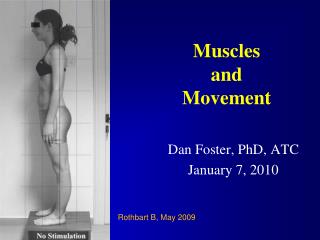
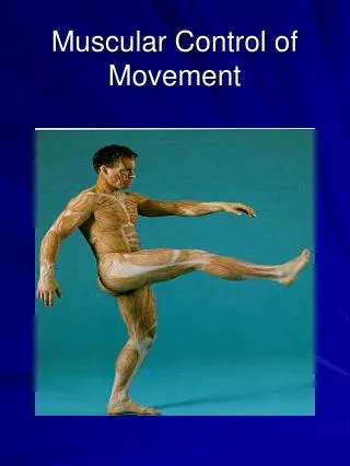
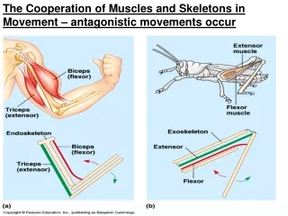
The Cooperation of Muscles and Skeletons in Movement – antagonistic movements occur
Thick Filaments: Thin Filaments: Myosin Actin Skeletal Muscle Nuclei Striations
Other Vertebrate Muscles • Smooth Muscle : • Lacks striations • Has less myosin than skeletal muscle • Found in walls of hollow organs (e.g. digestive tract organs) • Cardiac Muscle: • Structurally similar to skeletal muscle • Differs in action potential generation: • Action potentials spread throughout the heart through direct contact between cells
The Sliding-Filament Model of Muscle Contraction The length of the filaments stays the same as the muscle fiber contracts.
One Hypothesis for How Myosin-Actin Interactions Generate the Force for Muscle Contraction Myosin head is bound to ATP Binding a new molecule of ATP releases myosin from actin ATP -> ADP + P Releases ADP + P and returns to low-energy configuration
Regulatory protein that controls the position of tropomyosin Mechanism for the control of muscle contraction Regulatory protein that blocks myosin binding sites Ca ions bind to the troponin complex, which controls the position of tropomyosin.
The roles of the muscle fiber’s sarcoplasmic reticulum and T (Transverse) tubules in contraction Acetylcholine released infoldings of the plasma membrane a specialized ER
Each muscle fiber has a single synapse with one motor neuron, but each motor neuron typically synapses with several or many muscle fibers.
Motor Units in a Vertebrate Muscle Skeletal Muscle Fiber Axon Branch Axon Terminal