Download
1 / 35
350 likes | 487 Vues
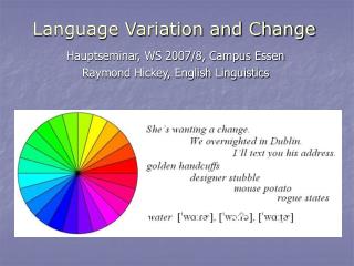
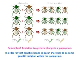
Variation forms the basis of evolution . There are two basic forms: 1 Continuous variation where individuals in a population shows a gradation from one extreme to the other. 2 Discontinuous variation where there is a limited number of distinct forms within the population.

E N D
Variation forms the basis of evolution. There are two basic forms: 1 Continuous variationwhere individuals in a population shows a gradation from one extreme to the other. 2 Discontinuous variation where there is a limited number of distinct forms within the population. Ch 10: Genetic Change and Variation
10.1.1 Table of data 10.1.2 Line graph 10.1 Methods of Recording Variation
10.1.3 Histogram 10.1.4 Bar graph
10.1.5 Kite graph 10.1.6 Pie chart
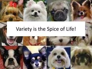
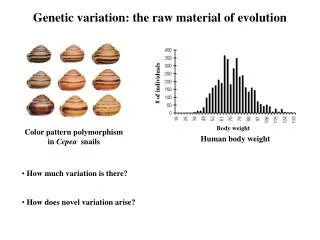
10.2.1 Continuous variation Characteristics within a population vary only very marginally between one individual and the next a graduation from one extreme to the other examples: weight, height, IQ, EQ, etc Characteristics which show continuous variation are controlled by the combined effect of a number of genes (polygenes) - a polygenic character 10.2 Types of Variation
The random assortment of genes during metaphase I of meiosis ensures that individuals possess a range of genes from any polygenic complex: all tall genes very tall all short genes very short about 1/2 tall and 1/2 short genes intermediate height 10.2 Types of Variation
The mean (arithmetic mean) is the average of a group of values. The mode is the single value of a group which occurs most often. The median is the central or middle value of a set of values. 10.2.2 The normal distribution curve
Thestandard deviation is a value which gives an indication of the range of values on either side of the mean.
characters which do not show a gradation between extremes but fall into a number of distinct forms usually controlled by a single gene which may have 2 or more alleles 10.3 The chi-squared test (not required in syllabus) 10.4 The t-test (not required in syllabus) 10.2.3 Discontinuous (discrete) variation
1. Environment 2. Genetic change: reshuffling of genes and mutation 10.5.1 Environmental effects Phenotype is the result of its genotype and effect of theenvironment. Because environmental influences are themselves very various and often form gradations, e.g. temperature, light intensity, etc., they are largely responsible for continuous variation within a population. 10.5 Origins of Variation
10.5.2 Reshuffling of genes - creating new combinations during sexual reproduction by: 1 Mixing two different parental genotypes where cross fertilization occurs 2 Random distribution of chromosomes during metaphase I of meiosis 3 Crossing over between homologous chromosomes during prophase I of meiosis 10.5 Origins of Variation
Mutationis any change in the structure or the amount of DNA of an organism. Most mutations occur in body cells and do not pass to offspring. Only those that affect gametes can be inherited and produce sudden and distinct differences between individuals discontinuous variation Mutation
Gene mutation(point mutation) is a changein the structure of DNA which occurs at a single locus on a chromosome gene mutation wrong sequence of amino acids no enzyme absence of a character, e.g. pigment 10.5.3 Changes in gene structure (point mutation)
There are many forms of gene mutation: 1. Duplication - a portion of the nucleotide chain becomes repeated 2. Addition (insertion) - an extra nucleotide sequence becomes inserted in the chain 3.Deletion – a portion of the nucleotide is removed from the chain 4. Inversion – a nucleotide sequence separates and rejoins at original position 5. Substitution – one of the nucleotides is replaced by another with a different base 10.5.3 Changes in gene structure (point mutation)
example: sickle-cell anaemia is the result of the replacement of just one base in the DNA molecule causing the wrong amino acid being joined into two of the polypeptide chains which make up the haemoglobin molecule. but the disease is resistant to malaria ! 10.5.3 Changes in gene structure (point mutation)
Polyploidy is the possession of more than 2 complete sets of chromosomes. e.g. triploid means 3 sets; tetraploid means 4 sets. Formation of tetraploid offspring: fertilization of diploid gametes or whole set of chromosomes doubles after fertilization Formation of triploid offspring: Fertilization of a diploid gamete with a normal haploid gamete 10.5.4Changes in whole sets of chromosomes
Autopolyploidy– polyploidy within the same species Autopolyploidy can be induced by colchicine (a chemical) which inhibits spindle formation and so prevents chromosomes separating during anaphase. Triploids are sterile because they cannot form complete homologous pairings. If, however, a hybrid has a chromosome number which is a multiple of the original chromosome number, a new fertile species is formed, e.g. wheat (n=42) is the cross between wild grass (n=14) and emmer wheat (n=28)
Allopolyploidy – A fertile species having a chromosome number which is a multiple of the original haploid number Allopolyploidy is rare in animals, but relatively common in plants, including many food plants, e.g. wheat, coffee, banana, sugar cane, apple, tomatoes, etc The polyploid variety often have advantages, e.g. large fruits, tomatoes have more vitamin C, etc.
Non-disjunction occurs when one of the homologous chromosomes (23 pairs) fails to segregate during meiosis, gametes formed have 22 & 24 chromosomes. This is often fatal. Down's syndrome : 47 chromosomes (+ extra 21st chromosome) Down's syndrome often occurs in ova formation rather than sperms, especially in old age pregnancies. 10.5.5 Changes in chromosome number
Turner's syndrome: Have one missing X chromosome XO with 45 chromosomes Females with small stature & sexually immature Klinefelter's syndrome: Genotypes are XXY, XXXY or XXXXY Males with small testes but no sperms, with breast development and female figures It indicates that Y is the cause of maleness
This occurs in meiosis when crossing over takes place. 1 Deletion 2 Inversion 3 Translocation 4 Duplication 10.5.6 Changes in chromosome structure
10.6 Causes of Mutations There is natural mutation rate which varies from one species to another. Animals with shorter life cycles show a greater rate of mutation because of more frequent meiosis. This natural mutation rate can be increased artificially by certain chemicals e.g. colchicine, formaldehyde, nitrous acid & mustard gas or energy sources (mutagens), e.g. uv rays, X rays, rays, & particles and neutrons
To risk for a mother to have babies with certain genetic diseases could be calculated, if enough information of the disease in the family is known, e.g. Down's syndrome, haemophilia. On the basis of this advice parents can choose whether or not to have children. Doctors can diagnose certain genetic defects, e.g. Down's syndrome, in a foetus, by studying samples of cells taken from the amniotic fluid which surrounds the foetus – a process called amniocentesis. Parents can then decide to have the pregnancy terminated. 10.7 Genetic Screening and Counselling –
10.7.1 Gene tracking To find out on which chromosome a defective gene is located. Blood groups are traced in families to act as gene markers. Correlation between certain blood groups alleles and the occurrence of a genetic disease can determine whether or not the gene for the disease is on the same chromosome as that for blood groups. If one genetic marker is not linked to the disease in question another must be tried and so on until the one which shows linkage with the disease is found. Linked markers are then used to work out if someone carries a disease.
19. 88-II-4 (b) What is the genetic basis of • Hybrid vigour, (5 marks) • and the determination of the ABO blood groups? (4 marks)