Download
1 / 19
210 likes | 418 Vues
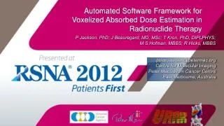
P Jackson, PhD; J Beauregard, MD, MSc; T Kron, PhD, DIPLPHYS; M S Hofman, MBBS; R Hicks, MBBS price.jackson@petermac.org Centre for Molecular Imaging Peter MacCallum Cancer Centre East Melbourne, Australia.

E N D
P Jackson, PhD; J Beauregard, MD, MSc; T Kron, PhD, DIPLPHYS; M S Hofman, MBBS; R Hicks, MBBS price.jackson@petermac.org Centre for Molecular Imaging Peter MacCallum Cancer Centre East Melbourne, Australia Automated Software Framework for Voxelized Absorbed Dose Estimation in Radionuclide Therapy
Purpose: • Accurately map distribution of radiation dose to healthy & target tissues in Peptide Receptor Radionuclide Therapy (PRRT) • Treatment given over 4 or more cycles • Opportunity to adjust prescription based on absorbed dose estimate? Small volume disease, limited absorption in lesion, high SUV in kidneys & spleen Bulky & highly octreotate-avid liver lesions, limited uptake observed in healthy organs
Internal Emitter Dosimetry • Radionuclide Dosimetry as function of: • Pharmacokinetics • Biological half-life, uptake & clearance (multiple phases) • Physical half-life • Emission particles • Type & energy • Absorbing medium • Generally soft-tissue (high-dose regions) • MIRD: OLINDA/EXM (Gold Standard) • Male/Female/child MIRD phantoms with customizable organ mass • Absorbed dose values accurate when considered over large population • Difficult to account for absorbed dose to tumour/target volume • Not intended for individualised dosimetry “NOTE: This code gives doses for stylized models of average individuals - results should be applied with caution to specific subjects.” Not an issue for short-range beta emitters, though • Significant Manual input • No accounting for sub-organ kinetics (renal pelvis vs. cortex)
Image Registration 4-Hour Fused SPECT/CT 24-Hour Fused SPECT/CT 72-Hour Fused SPECT/CT 4-Hour CT Fused to Cumulated Activity Image (Bq*Hr/mL) 4-Hour CT Fused to Absorbed Dose Image (mGy) Voxel S-Value & Gamma Convolution Kernel 4-Hour CT Fused to 4-, 24- & 72-hour SPECT Voxelized Kinetics Curve Fitting parameters per voxel Video: Interpolated activity & Cumulated activity Visual Analysis Voxelized Registration and Kinetics (VRAK) • In-house dosimetry protocol based on serial SPECT/CT: • Deformable Registration • Automatic Kinetics fitting & activity integration • Voxel S-Values (EGSnrc) • Output: dicom dose & cumulated activity volumes, data file with per voxel kinetics parameters • All scripted • Called from command-line w/text input file • Python-based (pydicom, numpy) • Open-source dependencies • Cross-platform (Win, Mac, Unix)
Quantitative SPECT CT: • 177Lu: 112 keV (6.2%) & 208.4 keV (10.4%) γ’s, 497 keVmax β- (100%) • 3 Quantitative SPECT/CT series • 4, 24, 72 hours Post-injection • SPECT quantitation previously calibrated* • 177Lu-specific attenuation & dead-time correction • 2 couch position (chest+abdomen) • Full datasets for 28 LuTate Rx’s (18 different patients) used for validation *Beauregard, J.-M. et al., 2011. Quantitative (177)Lu SPECT (QSPECT) imaging using a commercially available SPECT/CT system. Cancer imaging: the official publication of the International Cancer Imaging Society, 11, pp.56-66.
Image Registration : SPECT/CT Post-registration SPECT/CTs: • Sequential Rigid & Deformable • Elastix* (ITK-based, adjustable parameters, scriptable) • Multi-resolution B-Spline Deformation • Mutual Information Metric (80%) + deformation penalty (20%) • Robust, few ‘optical flow’-type artifacts • Co-register Anatomical (CT) volumes • Warp functional (SPECT) images • Inspect registered SPECTs • Apply translation where necessary • Output: 3 SPECT volumes aligned to 4- hour CT (fixed image) array space • Same resolution & origin (Image Position Patient 4-hour 24-Hour Difference/Overlay * Klein, S., Staring, M. & Pluim, J.P.W., 2007. Evaluation of optimization methods for nonrigid medical image registration using mutual information and B-splines. IEEE transactions on image processing : a publication of the IEEE Signal Processing Society, 16(12), pp.2879-90. 4-hour: Red 24-hour: Green
Aligned Images Voxelized Kinetics 72 Hr SPECT 24 Hr SPECT 4 Hr SPECT • 3 Co-registered SPECT Series 1 Volume (cumulated activity) • Read Time P.I. From DICOM header • Decay-correct activity values • Voxel-by-voxel • Analytical fit of 3-phase exponential pharmacokinetics • 1 Uptake, 2 Clearance • -A1=A2+A3 (C=0 at t=0) • K1>k2>k3 • Ignore values ~0 (out of patient or not relevant for dosimetry) • Adjust unrealistic, noisy values • Calculate for slope (as exponential) between measurements • Solve k3, A3 • Solve k2, A2 • Solve k1 • Limit Rate constants (k) to realistic range where necessary • Weighted for final time point • Single-threaded, but efficient (50-100x improvement over iterative curve-fitting routine: Scipy.Optimize) • 25M Voxels/Hr • 2-Position Chest/Abdo SPECT: 1.5 Hours Cumulated Activity
Kinetics Processing • 3x SPECT Array of kinetics parameters (A1, k1, A2, k2, A3, k3): [x,y,z,6] • Integrate decay-adjusted curves for disintegrations per unit volume • Output: Cumulated Activity (Bq*Hrs)/(mL) • Save as dicom (.dcm) or ITK (.mhd/.raw) file • Visual Output • Can be use to create image sequence of uptake & clearance • Arguments: slice #,projection (axial, sagittal, coronal), time window, # of frames • Save frames at times t • Interpolated Activity • Interpolated cumulated activity • Informative evaluation of relative uptake & clearance • Contribution of activity at time t to total # of disintegrations Activity t=0-100Hrs Cumulated Activity (Bq/mL) (Bq*Hr/mL)
Dose Calculation Point Spread Function EDKnrc Sphere Model Cumulated Activity Absorbed Dose • EGSnrc Simulation for beta- andphoton components of 177LuDecay • All tissue assumed to be H2O equivalent • Voxel S-Values for beta- component • Local energy deposition (only voxel self-dose) • Agreement with publishedS-values* • Long-range Gamma Voxel Dose Kernel • Low-resolution (high-efficiency) • 1.6*1.6*2.0 cm Voxels • Kernel calculated by dosxyz (EGSnrc) • Convolved through activity array VSV Calculation dosxyz (EGSnrc) *Lanconelli, N. et al., 2012. A free database of radionuclide voxel S values for the dosimetry of nonuniform activity distributions. Physics in medicine and biology, 57(2), pp.517-33.
Beta- Range vs. SPECT Resolution Range of beta electron transport shorter than range of SPECT Partial Volume effect SPECT Partial Volume effect for point source: Gaussian spread w/FWHM 7-15 mm* Absorbed Dose range for 177Lu beta-: 1-2 mm Activity heterogeneity clearly evident in PET \ Beta- energy from apparent activity (as seen on SPECT) considered to be deposited locally (in same voxel) 68Ga-Octreotate PET Imaging 1.1 Hrs PI * Gear, J. et al., 2011. Monte Carlo verification of polymer gel dosimetry applied to radionuclide therapy: a phantom study. Physics in Medicine and Biology, 56, p.7273. 177Lu-Octreotate SPECT Imaging 24 Hrs PI
Gamma Dose Convolved Cumulated Activity Array Gamma Kernel (dosxyz) • Small proportion of total dose • ~5-20% depending on region • Greater Range • Soft dose gradient • Dose Kernel from DOSxyz • Low-resolution (voxels 1.6*1.6*2.0 cm3) • Long range (21 cm max) • In processing: • Convolve through activity array • Combine with beta component for total absorbed dose • Output can be written as dose volume from beta-, gamma, combined • maintains alignment to CT data Gamma dose (13%) Beta- dose (87%)
Validation Methods • Coregister Serial SPECT/CT Volumes • Segment fixed CT volume in each study • Lower Large Intestine, Small intestine, Stomach, Upper Large Intestine, Heart , Kidneys, Liver, Lungs, Muscle, Pancreas, Marrow, Spleen, Bladder, Lesion • OLINDA Analysis • Apply segmentation to serial SPECT scans • Input mean organ activity values • Compute mean organ dose • VRAK Analysis • Apply segmentation to dose volume • Report Mean organ dose
Results: Cumulated Activity & Organ Dose as compared to OLINDA *Compartmental Organs in MIRD model (separate source & target regions; contents + wall) At Riskorgans (Somatostatin Analog PRRT)
Results: Cumulated Activity & Organ Dose as compared to OLINDA • Estimate of decays and total dose to kidneys, liver & spleen in the range of 5% with respect to conventional technique • Segmenting both contents and wall of compartmental organs overestimates dose by 30-100% • Greatest discrepancy in bladder • Similar effect observed in marrow • Comparable estimate of cumulated activity, but higher dose in segmented volume • Geometric effect accounted for by organ S-Value • Selective segmentation required • But susceptible to partial volume effect Compartmental Organ Model Source Volume Tissue (Bladder Wall) MIRD Bladder: Ellipsoid Source (Radius x,y,z: 4.71, 3.21, 3.21 cm) Outer Wall (Tissue) 2.5 mm thickness
Results: Organ S-Value • VRAK beta- VSV + gamma kernel closely matches MIRD S-values for solid organs • Common at risk organs in PRRT • Lesions are solid volumes too • Mean Lesion Dose (between all studies): 24.5 ± 9.7 Gy • ~10:1 ratio lesion to kidney dose (10.1±6.0) • Fraction of total patient dose from beta- emission: 87.1±0.75% • MIRD model better suited to compartmental organs • But MIRD schema does not account for self-dose to GI wall, only from contents • Relevant in somatostatin analog therapy
HU mGy 4-Hour CT VRAK Dose Discussion • Efficient, automated tool for PRRT dosimetry using serial functional images • Close agreement with OLINDA in both kinetics and dose estimates at organ level for 177Lu-octreotate dataset • Smooth, predictable kinetics estimation • No artifacts or noise from processing • Visually clear dosimetry data that can be overlaid with CT volume • Non-specific in design: can be applied to other isotopes, quantitative imaging (PET) • Need to know: physical half-life, isotope VSV & Gamma Kernel (dosxyz) • Personalized dosimetry data from initial cycle may be used to inform subsequent therapies • Radionuclide dosage • Extended renal-protective measures • Monitor relevant biochemical markers for at-risk organs
Discussion • Limitations: • Homogenous tissue (H2O) assumed • Resolution of SPECT (1-2 cm) • Detection of highly heterogeneous uptake? • Disintegrations in tumour spread across larger volume • Reduces estimate of absorbed dose in lesion • Registration accuracy varies • In range of 5 mm for most organs • Other algorithms (Demons, etc) can be more precise, but prone to artifacts • Inconsistent breath hold can shift activity near diaphragm (liver & metastases, spleen) • Kinetics Fitting may overestimate cumulated activity to organs with transient uptake at early time point (bladder, bowel) • Not observed in critical organs, tumour volumes Anatomical Volume Approx. Margin of SPECT volume
Project Homepage • VRAK: http://code.google.com/p/vrak-dosimetry/ • Dependencies: • Elastix (http://elastix.isi.uu.nl/about.php) • Python 2.7 (http://www.python.org/download/releases/2.7.3/) • Pydicom (0.9.7) (http://code.google.com/p/pydicom/downloads/list) • Numpy (1.6.2) (http://sourceforge.net/projects/numpy/files/) • For Video: • ffmpeg (compiled from source) (http://ffmpeg.org/download.html) • PIL (http://www.pythonware.com/products/pil/) • Matplotlib (1.1.1) (http://sourceforge.net/projects/matplotlib/files/matplotlib/matplotlib-1.1.1/) • Recommended Viewer: 3D Slicer (http://www.slicer.org/)
References • Beauregard, J.-M. et al., 2011. Quantitative (177)Lu SPECT (QSPECT) imaging using a commercially available SPECT/CT system. Cancer imaging: the official publication of the International Cancer Imaging Society, 11, pp.56-66. • Lanconelli, N. et al., 2012. A free database of radionuclide voxel S values for the dosimetry of nonuniform activity distributions. Physics in medicine and biology, 57(2), pp.517-33. • ICRP, 1983. Radionuclide Transformations: Energy and Intensity of Emissions. In ICRP Publication 38. Pergamon Press. • Kawrakow, I. & Walters, B.R.B., 2006. Efficient photon beam dose calculations using DOSXYZnrc with BEAMnrc. Medical Physics, 33(8), p.3046. • Klein, S., Staring, M. & Pluim, J.P.W., 2007. Evaluation of optimization methods for nonrigid medical image registration using mutual information and B-splines. IEEE transactions on image processing: a publication of the IEEE Signal Processing Society, 16(12), pp.2879-90. • Stabin, M.G., Sparks, R.B. & Crowe, E., 2005. OLINDA/EXM: the second-generation personal computer software for internal dose assessment in nuclear medicine. Journal of nuclear medicine: official publication, Society of Nuclear Medicine, 46(6), pp.1023-7. • Gear, J. et al., 2011. Monte Carlo verification of polymer gel dosimetry applied to radionuclide therapy: a phantom study. Physics in Medicine and Biology, 56, p.7273. • Eckerman, K. et al., 1994. Availability of nuclear decay data in electronic form, including beta spectra not previously published. Health physics, 67(4), pp.338-345.