Download
1 / 72
780 likes | 1.17k Vues
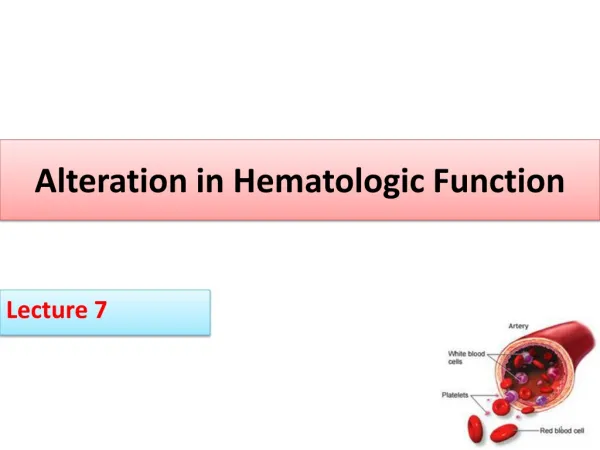
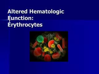
Alterations of Hematologic Function. Chapter 20. Anemia. Reduction in the total number of erythrocytes in the circulating blood or in the quality or quantity of hemoglobin Impaired erythrocyte production Acute or chronic blood loss Increased erythrocyte destruction Combination of the above.

E N D
Alterations of Hematologic Function Chapter 20
Anemia • Reduction in the total number of erythrocytes in the circulating blood or in the quality or quantity of hemoglobin • Impaired erythrocyte production • Acute or chronic blood loss • Increased erythrocyte destruction • Combination of the above
Anemia • Classifications • Etiology • Morphology • Based on MCV, MCH, and MCHC values • Size • Identified by terms that end in “-cytic” • Macrocytic, microcytic, normocytic • Hemoglobin content • Identified by terms that end in “-chromic” • Normochromic and hypochromic
Anemia • Anisocytosis • Red cells are present in various sizes • Poikilocytosis • Red cells are present in various shapes
Anemia • Physiologic manifestation • Reduced oxygen-carrying capacity • Variable symptoms based on severity and the ability for the body to compensate • Classic anemia symptoms • Fatigue, weakness, dyspnea, and pallor
Macrocytic-Normochromic Anemias • Also termed megaloblastic anemias • Characterized by defective DNA synthesis • Caused by deficiencies in vitamin B12 or folate • Coenzymes for nuclear maturation and the DNA synthesis pathway
Macrocytic-Normochromic Anemias • Ribonucleic acid (RNA) processes occur at a normal rate • Results in the unequal growth of the nucleus and cytoplasm
Macrocytic-Normochromic Anemias • Pernicious anemia • Caused by a lack of intrinsic factor from the gastric parietal cells • Required for vitamin B12 absorption • Results in vitamin B12 deficiency
Macrocytic-Normochromic Anemias • Pernicious anemia • Typical anemia symptoms • Neurologic manifestations • Nerve demyelination • Absence of intrinsic factor • Others • Loss of appetite, abdominal pain, beefy red tongue (atrophic glossitis), icterus, and splenic enlargement
Macrocytic-Normochromic Anemias • Pernicious anemia • Treatment • Parenteral or high oral doses of vitamin B12
Macrocytic-Normochromic Anemias • Folate deficiency anemia • Absorption of folate occurs in the upper small intestine • Not dependent on any other factor • Similar symptoms to pernicious anemia except neurologic manifestations generally not seen • Treatment requires daily oral administration of folate
Microcytic-Hypochromic Anemias • Characterized by red cells that are abnormally small and contain reduced amounts of hemoglobin • Related to: • Disorders of iron metabolism • Disorders of porphyrin and heme synthesis • Disorders of globin synthesis
Microcytic-Hypochromic Anemias • Iron deficiency anemia • Most common type of anemia worldwide • Nutritional iron deficiency • Metabolic or functional deficiency • Progression of iron deficiency causes: • Brittle, thin, coarsely ridged, and spoon-shaped nails • A red, sore, and painful tongue
Microcytic-Hypochromic Anemias • Sideroblastic anemia • Group of disorders characterized by anemia • Altered mitochondrial metabolism causing ineffective iron uptake and resulting in dysfunctional hemoglobin synthesis • Ringed sideroblasts within the bone marrow are diagnostic • Sideroblasts are erythroblasts that contain iron granules that have not been synthesized into hemoglobin
Microcytic-Hypochromic Anemias • Sideroblastic anemia • Dimorphism • Myelodysplastic syndrome • Erythropoietic hemochromatosis
Normocytic-Normochromic Anemias • Characterized by red cells that are relatively normal in size and hemoglobin content but insufficient in number
Normocytic-Normochromic Anemias • Aplastic anemia • Pancytopenia • Pure red cell aplasia • Fanconi anemia • Posthemorrhagic anemia • Acute blood loss from the vascular space
Normocytic-Normochromic Anemias • Hemolytic anemia • Accelerated destruction of red blood cells • Autoimmune hemolytic anemias • Immunohemolytic anemia • Warm antibody immunohemolytic anemia • Drug-induced hemolytic anemia • Cold agglutinin immunohemolytic anemia • Cold hemolysin hemolytic anemia
Normocytic-Normochromic Anemias • Sickle cell anemia • Anemia of chronic inflammation • Mild to moderate anemia seen in: • AIDS, rheumatoid arthritis, lupus erythematosus, hepatitis, renal failure, and malignancies
Normocytic-Normochromic Anemias • Anemia of chronic inflammation • Pathologic mechanisms • Decreased erythrocyte life span • Ineffective bone marrow response to erythropoietin • Altered iron metabolism
Myeloproliferative RBC Disorders • Polycythemia • Overproduction of red blood cells • Relative polycythemia • Result of dehydration • Fluid loss results in relative increases of red cell counts and Hgb and Hct values
Polycythemia • Absolute polycythemia • Primary absolute • Abnormality of stem cells in the bone marrow • Polycythemia vera (PV) • Secondary absolute • Increase in erythropoietin as a normal response to chronic hypoxia or an inappropriate response to erythropoietin-secreting tumors
Alterations of Leukocyte Function • Quantitative disorders • Increases or decreases in cell numbers • Bone marrow disorders or premature destruction of cells • Response to infectious microorganism invasion • Qualitative disorders • Disruption of cellular function
Quantitative Alterations of Leukocytes • Leukocytosis • Leukocytosis is a normal protective physiologic response to physiologic stressors • Leukopenia • Leukopenia is not normal and not beneficial • A low white count predisposes a patient to infections
Granulocytosis (Neutrophilia) • Neutrophilia is evident in the first stages of an infection or inflammation • If the need for neutrophils increases beyond the supply, immature neutrophils (banded neutrophils) are released into the blood
Granulocytosis (Neutrophilia) • This premature release is detected in the manual WBC differential and is termed a shift to the left • When the population returns to normal, it is termed a shift to the right
Neutropenia • Reduction in circulating neutrophils • Causes • Prolonged severe infection • Decreased production • Reduced survival • Abnormal neutrophil distribution and sequestration
Granulocytopenia (Agranulocytosis) • Causes • Interference with hematopoiesis • Immune mechanisms • Chemotherapy destruction • Ionizing radiation
Eosinophilia • Hypersensitivity reactions trigger the release of eosinophilic chemotactic factor of anaphylaxis from mast cells • Increased in allergic disorders • Increased in parasitic invasions
Eosinopenia • Decrease in circulation numbers of eosinophils • Usually caused by migration of cells to inflammatory sites • Other causes • Surgery, shock, trauma, burns, or mental distress
Basophils • Basophils account for only 0% to 1% of the circulating WBCs • Basophilia • Response to inflammation and hypersensitivity reactions • Basopenia • Occurs in acute infections, hyperthyroidism, and long-term steroid therapy
Monocytes • Monocytosis • Poor correlation with disease • Usually occurs with neutropenia in later stages of infections • Monocytes are needed to phagocytize organisms and debris • Monocytopenia • Very little known about this condition
Lymphocytes • Lymphocytosis • Acute viral infections • Epstein-Barr virus • Lymphocytopenia • Immune deficiencies, drug destruction, viral destruction
Infectious Mononucleosis • Acute, self-limiting infection of B-lymphocytes transmitted by saliva through personal contact • Commonly caused by the Epstein-Barr virus (EBV)—85% • B cells have an EBV receptor site • Others viral agents resembling IM • Cytomegalovirus (CMV), hepatitis, influenza, HIV
Infectious Mononucleosis • Symptoms: fever, sore throat, swollen cervical lymph nodes, increased lymphocyte count, and atypical (activated) lymphocytes • Serious complications are infrequent (<5%) • Splenic rupture is the most common cause of death
Infectious Mononucleosis • >50% lymphocytes and at least 10% atypical lymphocytes • Diagnostic test • Monospot qualitative test for heterophilic antibodies • Treatment: symptomatic
Leukemias • Malignant disorder of the blood and blood-forming organs • Excessive accumulation of leukemic cells • Acute leukemia • Presence of undifferentiated or immature cells, usually blast cells • Chronic leukemia • Predominant cell is mature but does not function normally
Leukemias • Acute lymphocytic leukemia (ALL) • Acute myelogenous leukemia (AML) • Chronic myelogenous leukemia (CML) • Chronic lymphocytic leukemia (CLL)
Leukemias • Signs and symptoms of leukemia • Anemia, bleeding purpura, petechiae, ecchymosis, thrombosis, hemorrhage, DIC, infection, weight loss, bone pain, elevated uric acid, and liver, spleen, and lymph node enlargement
Lymphadenopathy • Enlarged lymph nodes that become palpable and tender • Local lymphadenopathy • Drainage of an inflammatory lesion located near the enlarged node • General lymphadenopathy • Occurs in the presence of malignant or nonmalignant disease
Malignant Lymphomas • Malignant transformation of a lymphocyte and proliferation of lymphocytes, histiocytes, their precursors, and derivatives in lymphoid tissues • Two major categories • Hodgkin lymphoma • Non-Hodgkin lymphomas
Hodgkin Lymphoma • Characterized by the presence of Reed-Sternberg cells in the lymph nodes • Reed-Sternberg cells are necessary for diagnosis, but they are not specific to Hodgkin lymphoma • Classical Hodgkin lymphoma • Nodular lymphocyte predominant Hodgkin lymphoma
Hodgkin Lymphoma • Physical findings • Adenopathy, mediastinal mass, splenomegaly, and abdominal mass • Symptoms • Fever, weight loss, night sweats, pruritus • Laboratory findings • Thrombocytosis, leukocytosis, eosinophilia, elevated ESR, and elevated alkaline phosphatase • Paraneoplastic syndromes
Non-Hodgkin Lymphoma • Generic term for a diverse group of lymphomas • The lymphomas can be differentiated based on etiology, unique features, and response to therapies