Adrenocortical carcinoma
660 likes | 1.22k Vues
Adrenocortical carcinoma. Presented by: Dr. M. K. M. Foisal Quader Chowdhury Chittagong Medical College Hospital. Case presentation . Name of the patient: Md. Yunus . Age: 30 years. Address: Puichori , Banskhali . Occupation: Shopkeeper.

Adrenocortical carcinoma
E N D
Presentation Transcript
Adrenocortical carcinoma Presented by: Dr. M. K. M. Foisal Quader Chowdhury Chittagong Medical College Hospital
Case presentation Name of the patient: Md. Yunus. Age: 30 years. Address: Puichori, Banskhali. Occupation: Shopkeeper. Date of admission: 20th August, 2013. Bed: 09, Ward: 24,Reg: 7324/ 89. CMCH.
Case presentation Chief Complaints: • Generalized weakness, weight loss, effort intolerance, occasional vertigo, burning sensation in the scalp for 8 months; • Anorexia for 6 months, • Diffuse pain in the right flank and right upper abdomen, grasping in nature associated with frequent rise of temperature for 4 months; • Urgency with Frequency of Micturition and occasional sleep disturbances for 2 months.
Case presentation For these complaints he consulted local doctor and was advised for ultrasonograph of whole abdomen that revealed (24 – 06 – 13) big solid retroperitoneal mass above right kidney, later, from which, ultrasonograph guided FNAC was taken that revealed (29 – 06 – 13) Adrenocortical carcinoma. Then he was referred to CMCH for further management. Patient is non diabetic, non hypertensive, non icteric, with no history of any other major medical or surgical illness.
Case presentation General examination: On admission, findings were almost within normal range with Pulse 72 / min; Blood pressure 90 / 60 mm of Hg; Not anaemic, cyanosed, edematous or dehydrated; No abnormality was detected on examination of chest with normal heart sound & clear, vesicular breath sound. No enlarged lymph node was found on palpation.
Case presentation Local Examination: His abdomen was soft, not distended, No palpable organomegally or intra-abdominal mass, No scar mark, Hernial orifice was intact, Mild tenderness in right hypochondriac region and right renal angle. His bowel and bladder habit has also been normal.
Case presentation His investigation profile shows :
Case presentation His investigation profile shows :
Case presentation His investigation profile shows :
Case presentation On the basis of the clinical presentation and investigation profile, the patient was diagnosed as a case of Adrenocortical Carcinoma of non functioning variety.He has been on conservative management so far. Next plan for management was Surgical resection of the tumor after senior consultation. In order to prepare for the perioperative medical, hormonal & other possible complications, a medical board was arranged on 31st August, 2013.
Case presentation Medical Board: Date: 31 – 08 – 13 ; Time: 10:00 AM ; Ward: 24. Respected members of Medical Board: • Professor Dr. Omar Faruque Yusuf, Head, Dept. of Surgery. • Professor Dr. A. K. M. ShamsulAlam, Head, Dept. of Anesthesiology. • Professor Dr. Md. MahtabUddinHasan, Head, Dept. of Medicine. • Professor Dr. Khandker A. K. Azad, Hesd, Surgery unit – II • Associate Professor Dr. G. M. JakirHossain, Dept. of Surgery. • Associate Professor Dr. IftekharHossain Khan, Dept. of Endocrinology.
Case presentation Medical Board: Conclusion: The respected members of Medical Board have examined the patient and gone through all the papers and diagnosed the patient as a case of Adrenocortical Carcinoma. Detailed discussion regarding hormonal evaluation, possible perioperative complications and necessary precaution & preparation for their management took place. Finally, following decisions were made: • Surgical resection of the Tumor; • Special Precaution for the HBsAg; • Essential support for the hormonal fluctuation and other medical emergencies during and after operation.
DISCUSSION On Adrenocortical Carcinoma
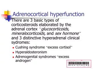
Physiology Adrenal Medulla secrets Epinephrine and nor-epinephrin. • Aldosteron = Helps to maintain water & electrolyte balance, by absorption of Na+ & excretion of K+. • Cortisol = Helps to maintain blood gloucose level by affecting glucose metabolism in the body. • Androgen = Promotes Hair growth, masculinization. • Oestrogen = Promotes feminization. • Epinephrine and nor-epinephrine = Stimulates body’s sympathetic activity.
Tumors of Adrenal Gland Non-functional and Benign: • Adenoma • Myelolipoma • Hematoma • Cyst • MacronodularHyperplasia
Tumors of Adrenal Gland Functional tumors: • Adrenal Medullary Tumors: • Pheochromocytoma • Childhood tumors: ganglioneuromas, neuroblastomas • Adrenal Cortical Tumors: • Cortisol-secreting adenoma • Aldosteronoma • Adrenocortical Carcinoma
Types of Adrenocortical Ca • Oncocyticadrenal cortical carcinoma, • Myxoidadrenal cortical carcinoma, • Carcinosarcoma, • Adenosquamousadrenocortical carcinoma, • Clear cell adrenal cortical carcinoma. • Secondary metastatic tumor of adrenal gland.
Etiology & genetics of Adrenocortical Carcinoma Risk factors for adrenocortical carcinoma include having the following hereditary diseases: • Li-Fraumeni syndrome. • Beckwith-Wiedemann syndrome. • Carney complex. Following genes have been found to be involved: • IGF-II gene • Suppression of p53 gene • c-myc gene
Staging of Adrenocortical Carcinoma The following stages are used for adrenocortical carcinoma: Pea, peanut, walnut, and lime show tumor sizes.
Staging of Adrenocortical Carcinoma According to the International Union Against Cancer: Tumor criteria T1 - Tumor diameter £ 5 cm with no local invasion T2 - Tumor diameter > 5 cm with no local invasion T3 - Tumor of any size with local extension but not involving adjacent organs T4 - Tumor of any size with local invasion of adjacent organs Lymph node criteria N0 - No regional lymph node involvement N1 - Positive regional nodes Metastasis criteria M0 - No distant metastasis M1 - Distant metastasis
Staging of Adrenocortical Carcinoma Stages Stage 1 - T1, N0, M0 Stage 2 - T2, N0, M0 Stage 3 - T1 or T2, N1, M0 T3, N0, M0 Stage 4 - T3, N1, M0 T4, any N, M0 Any T, any N, M1
Clinical presentation of Adrenocortical Carcinoma A functioning adrenocortical tumor makes too much of one of the following hormones: • Cortisol. • Aldosterone. • Testosterone. • Estrogen.
Clinical presentation of Adrenocortical Carcinoma Too much cortisol may cause: • Weight gain in the face, neck, and trunk of the body and thin arms and legs. • Growth of fine hair on the face, upper back, or arms. • A round, red, full face (Moon face). • A lump of fat on the back of the neck ("buffalo hump”). • A deepening of the voice and swelling of the sex organs or breasts in both males and females. • Muscle weakness. • High blood sugar. • High blood pressure.
Clinical presentation of Adrenocortical Carcinoma Too much aldosterone may cause: • High blood pressure. • Muscle weakness or cramps. • Frequent urination. • Feeling thirsty. Too much testosterone (in women) may cause: • Growth of fine hair on the face, upper back, or arms. • Acne. • Balding. • A deepening of the voice. • No menstrual periods.
Clinical presentation of Adrenocortical Carcinoma Too much estrogen (in women) may cause: • Irregular menstrual periods in women who have not gone through menopause. • Vaginal bleeding in women who have gone through menopause. • Weight gain. Too much estrogen (in men) may cause: • Growth of breast tissue. • Lower sex drive. • Impotence.
Clinical presentation of Adrenocortical Carcinoma • Adrenocortical carcinoma may present differently in children and adults. Most tumors in children are functional, and virilization is by far the most common (~80%) presenting symptom, followed by Cushing's syndrome (~6%) and precocious puberty. • Among adults presenting with hormonal syndromes, Cushing's syndrome (~40%) alone is most common, followed by mixed Cushing's and virilization (glucocorticoid and androgen overproduction).
Clinical presentation of Adrenocortical Carcinoma • Feminization and Conn syndrome (mineralocorticoid excess) occur in less than 10% of cases. • Non-functional tumors (about 40%) usually present with abdominal or flank pain, or they may be asymptomatic and detected incidentally.
Clinical presentation of Adrenocortical Carcinoma Endocrine syndromes associated with adrenocortical carcinoma • Cushing syndrome (30%) • Virilization and precocious puberty (22%) • Feminization (10%) • Primary hyperaldosteronism (2.5%) • Combined hormone excess (35%) • Polycythemia ( < 1%) • Hypercalcemia ( < 1%) • Hypoglycemia ( < 1%) • Adrenal insufficiency (particularly from primary adrenal lymphomas) • Non–glucocorticoid-mediated insulin resistance • Catecholamine excess due to rare instances of coexisting pheochromocytoma • Cachexia (usually preterminal)
Differential Diagnosis Differential diagnosis includes: • Adrenocortical adenoma, • Renal cell carcinoma, • Adrenal medullary tumors, • Hepatocellular carcinoma. • Neuroblastoma. • Pancreatic Cancer.
Investigations Routine Investigation: • Complete Blood Count • Urine R/M/E • Chest X ray • ECG • Serum Creatinine • HBsAg • RBS/ FBS with 2 hrs post prandial blood sugar
Investigation Other tests for diagnosis: • USG of whole abdomen & USG guided FNAC. [In this case, this procedure first detected the pathology] • Biopsy – for confirmation of diagnosis and histological typing of cancer – done after surgical resection.
Investigation For staging: • CT scan • MRI • CT scans of the chest and bone scans are routinely performed to look for metastases to the lungs and bones respectively. These studies are critical in determining whether or not the tumor can be surgically removed. They are useful for identifying the site of the tumor, differentiating it from other diseases, such as adrenocortical adenoma, and determining the extent of invasion of the tumor into surrounding organs and tissues.
Treatment The only curative treatment is complete surgical excision of the tumor, which can be performed even in the case of invasion into large blood vessels, such as the renal vein or inferior vena cava. Surgical Care • Preoperative screening • Include a full evaluation to determine the extent of disease and staging, which has implications for the ultimate prognosis. • Look for the most common sites for metastases. They are the lungs, liver, bone, and lymph nodes. Contiguous spread to the kidney and liver (if the primary is on the right side) and tumor extension into the venous drainage system of the adrenals and the inferior vena cava are common.
Treatment Surgical resection When feasible, total resection remains the management modality of choice for the definitive management of AC. It also remains the only potentially curative therapeutic modality. Recurrent local and metastatic disease is common, even among patients who undergo a successful complete resection. In such settings, the only effective treatment is attempted reoperation.
Treatment Preoperative diagnostic accuracy should improve in the following years with improved MRI technology, percutaneous core needle biopsy technology, and advances in molecular, genetic, and immunotyping interpretation. Aggressive resection of locally recurrent disease may prolong survival in some patients, while percutaneous radiofrequency ablation (RAF) may have a place in providing local symptom control related to local compression by an invasive tumor.
Treatment Open Adrenalectomy Suitable for large tumor, esp when there is evidence/ suspicion for local spread.
Treatment Laparoscopic Adrenalectomy Suitable for smaller tumor, esp when there is no evidence/ suspicion for local spread. Ports for Laparoscopic Adrenalectomy
Treatment Role of radiotherapy: For a long time, radiotherapy seemed to be less effective for curative purpose. However, There are emerging case reports demonstrating improved outcomes when palliative XRT used for localized lesions.
Treatment Role of Chemotherapy: Mitotane • This drug remains the major chemotherapeutic option for the management of AC because it is a relatively specific adrenocorticalcytotoxin. It is used as primary therapy, as adjuvant therapy, and as therapy in recurrent or relapsing disease. • Chemical relative to DDT • Found to have adrenolytic activity in dogs in vivo (selectively destroyed the zonaereticularis and fasciculata)
Treatment Role of Chemotherapy: Mitotane • Side effects are major and frequent, including: • CNS disturbance (vertigo, somnolence, ataxia) • Liver Toxicity • Renal Toxicity • Nausea, Vomiting • Diarrhea • Rash
Treatment Role of Chemotherapy: Other symptom- palliative Options: • Metyrapone (11B hydroxylase inhibitor) • Ketoconazole • Aminoglutehamide Cytotoxics Most studied have been Etoposide, cisplatin, and adriamycin.
Treatment Newer options: Biologic therapy is a treatment that uses the patient's immune system to fight cancer. Substances made by the body or made in a laboratory are used to boost, direct, or restore the body's natural defenses against cancer. This type of cancer treatment is also called biotherapy or immunotherapy. Targeted therapy is a type of treatment that uses drugs or other substances to identify and attack specific cancer cells without harming normal cells.