Download
1 / 2
20 likes | 179 Vues
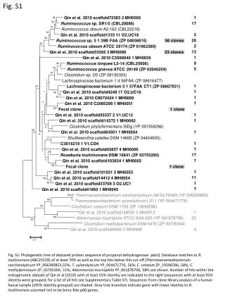
M. 1. 2. A. B. 8. 5. 6. 4. 7. 10. 10. 10. 10. 10. Isolation and Identification of Entomopathogenic Fungus against the pine sawyer, Monochamus saltuarius Gebler ( Coleoptera : Cerambycidae ). Days on 100% mortality. 600bp. P1-P3. 500bp. 400bp. P1-P5. 300bp. 200bp.

E N D
M 1 2 A B 8 5 6 4 7 10 10 10 10 10 Isolation and Identification of Entomopathogenic Fungus against the pine sawyer, MonochamussaltuariusGebler (Coleoptera: Cerambycidae) Days on 100% mortality 600bp P1-P3 500bp 400bp P1-P5 300bp 200bp conidia/ml Jeong-Mi Oh1, Tae-Young Shin1, Jae-Kyung Lee1, Hyun-Na Koo1, Soo-Dong Woo1 1Department of Agricultural Biology, Chungbuk National University, Cheongju, 363-763, Korea (sdwoo@cbnu.ac.kr) ABSTRACT Pine wilt is the most important disease of pine trees in Korea, Japan and China. The pathogen causing this disease, the pinewood nematode (Bursaphelenchus xylophylus), is transmitted vectored by adults of some cerambycid beetle species and the Japanese pine sawyer, Monochamus alternatus, is the major vector species in Korea. Although chemical insecticides have been used to kill vector insect and thus prevent transmission of the pathogen, the efficacy is not good. In Japan, to control this insect, an entomopathogenic fungus was studied and developed as an insecticide. This is thought to be the convenient and effective method to control M. alternatus. Recently, there are several reports about the pinewood nematode is vectored by also the pine sawyer, M. saltuarius, in Korea. The objective of this study, therefore, was to isolate and identify entomopathogenic fungi from to control it. We collected M. saltuarius cadaver the cadaver of M. saltuarius and then screened several fungi colonies. The pathogenicity of each fungus was tested using oak longicorn beetle, Moechotypa diphysis, as substitutive insect. M. diphysis is also serious pest to various trees in forest. As the result, only one of them showed high pathogenicity against M. diphysis. Selected fungus was identified by microscopic examination and DNA analysis. Pathogenicity was also evaluated to M. saltuarius 5’-AAGCTTCGACATGGTCTG–3’ P1 524 333 0 P3 5‘-GGAGGTGGTGAGGTTCTGTT-3’ P5 5‘-AGGAGAGAGCTCGACGGTCA-3’ Fig. 2. Phase contrast (A) and scanning electron (B) micrographs of MsW1. Conidia and mycelia were mounted from plates and examined under X400 on a Nikon microscope. For the SEM, samples were mounted on stubs, coated with gold and observed in a Carl Zeiss LEO-1530 SEM. Fig. 5. Location and sequence of PCR primers for differentiation of the Beauveria spp. Identification of enotomopathogenic fungi was conducted by previous reported primers, P1 (forward), P3 (backward), and P5 (backward) (Dwayne et al. 1996). PCR with the P1-P3 primer set could discriminate isolates of Beauveria spp from other entomopathogenic fungi with a PCR product of about 500bps. The P1-P5 primer set could identify B. bassiana strain specifically from Beauveria spp. with a approximately 330bp within P1-P3 PCR product. MsW1 was characterized by the sympodial development of single-celled conidia (ameroconidia) on a geniculate or zig-zag rachis. MATERIALS AND METHODS 1. lsolation of fungi Fungi were isolated directly from cadavers supporting fungal sporulation, using a semi-selective medium consisting of potato dextrose agar (PDA; DifcoTM, USA) containing PENICILLIN-STREPTOMYCIN SOLUTION (Sigma, USA). 2. Culture condition Fungi were grown on potato dextrose agar (PDA; DifcoTM, USA) in the dark at 25℃ and store at 4℃ until use. 3. Insects Adults M. saltuarius and Moechotypa diphysis were field-collected and used to bioassay. Fig. 3. Pathogenicity of MsW1 against to Moechotypa diphysis adults. The pathogenicity was evaluated as till times that mortality reaches to 100%. Conidia of MsW1 obtained from 14-day-old PDA plate were suspended in 0.02% Tween-80 solution. Spores were prepared from 1 × 10⁸ conidia/ml to 1 × 10⁴ conidia/ml by diirect counting in a haemocytometer. M. diphysis were inoculated by dipping for 10~15 sec into the spore suspension and were maintained in a plant culture chamber at 25℃. Fig. 6. Agarose gel electrophoresis of P1-P3 and P1-P5 PCR products from MsW1. Genomic DNA was extracted using an I-genomic BYF DNA extraction mini Kit (Intron Co., Korea). The reaction parameters for P1-P3 primer set were as follows; initial denaturation for 3 min at 94 ℃, followed by 35 cycles of 94℃ for 30sec, 53 ℃ for 30sec, and 72 ℃ for 45sec, and a final 10-min extension at 72 ℃. The reaction parameters for P1-P5 primer set were same with those of P1-P3 except the annealing temperature of 50 ℃. Lane M, 100 bp size marker; lane 1, P1-P3 PCR ; lane 2, P1-P5 PCR. RESULTS AND DISCUSSIONS Mortality of M. diphysis showed 100% at all concentarions and the pathogenicity increased with increasing conidial concentrations. The result of PCR with P1-P3 primer set indicated that MsW1 is belonged to Beauveria spp. To discriminate the species of MsW1 more precisely, PCR was performed with the P1-P5 primer set. As the result, a specific PCR product of about 330bp was amplified. This suggests that MsW1 is B. bassiana. B A B A CONCLUSIONS • Entomopathogenic fungus, MsW1, was isolated from • M. saltuarius and was identified with B. bassiana. • The pathogenicity of MsW1 against Moechotypa • diphysis, as substitutive insect, showed 100% • mortality at the tested all concentrations of conidia. • We are investigating the potential of MsW1 as a • biocontrol agent for M. saltuarius. Fig. 1. Selection of fungus from cadaver of M. saltuarius. Growth of the selected fungus on PDA medium at 25℃ for 3days (A) and 14days (B). REFERENCES Several fungal colonies were isolated from the cadaver and their pathogenicities were assayed against to Moechotypa diphysis for the selection of entomopathogenic fungus. As a result, a kind of fungus was obtained and named as MsW1. Colonies of MsW1 was usually slow growing, downy, at first white but later often becoming yellow to pinkish. Fig. 3. Moechotypa diphysis adults infected with MsW1 at 5 days (A) and 10 days (B) after death. The pathogenicity of MsW1 was also confirmed against M. saltuarius adults, but it needs more in detail and repeatedly. • Dwayne et. al. 1996. J. Invertebr. Pathol. 67:289-299. • Mitsuaki Shimazu. 2004.J. Appl. Entomol. 39(3):485-490
M1 M 1 2 3 4 5 958bp 1kbp - 45kDa - 34kDa - - - 30kDa 31kDa 26.5kDa - 19.5kDa - Introduction Methods Results (B) (A) MbPol-F primer 5’-ATG CGC TGG TAT AAT TTA GAA GAT -3’ Polyhedrin Gene 5’-CATG TCA ATA AAA TAC TTA CGT ATC A-3’ MbPol-R primer (B) (A) Conclusions References Pathogenicity and Molecular Characterization of nucleopolyhedrovirus isolated from Mamestra brassicae (Lepidoptera: Noctuidae) in Korea. Jeong-Mi Oh1, Jae-Kyung Lee1, Hyun-Na Koo1, Yeon-Ho Je2, Byung-Rae Jin3, Soo-Dong Woo1 1Department of agricultural Biology, Chungbuk National University, Cheongju, Korea, sdwoo@cbnu.ac.kr, 2School of Agricultural Biotechnology, Seoul National University, Seoul, Korea, and 3 College of Natural Resources and Life Science, Dong-A University, Busan, Korea 107 106 105 104 No. of dead larvae The cabbage armyworm, Mamestra brassicae, is an important insect pest of vegetables and ornamental plants in Asia and Europe. Several nucleopolyhedroviruses (NPVs) have been isolated from M. brassicae and considered useful biological-control agents for M. brassicae. However, Korean strain of M. brassicae NPV (MbNPV) is poorly characterized. For the practical use of NPV as a control agent of noctuid pests, it is necessary to identify and characterize the virus strain in advance. The objective of our study was the pathogenic, morphologic and genetic characterization of a MbNPV isolate (MbNPV-K1) derived from a diseased larva of M. brassicae found in Korea. 107 106 105 104 Fig. 5. Pathogenicity of MbNPV-K1(A) and Mamestrin(B) against 3rd instar larvae of M. brassicae. No. of dead larvae Fig. 3. SDS-PAGE analysis of the polyhedral proteins. p. i. days Larvae of M. brassicae were inoculated by a modified droplet-feeding method. Experiments were replicated three times with 25 larvae per treatment. Larvae were observed daily for mortality until 20 days after inoculation. Proteins from the polyhedra were subjected to a 12% SDS-PAGE and stained with Coomassie brilliant blue. Lane 1, polyhedrin of MbNPV-K1; lane 2, polyhedrin of Mamestrin; lane 3, polyhedrin of Autographa californica NPV; lane 4, polyhedrin of Spodoptera exigua NPV; lane 5, polyhedrin of Bombyx mori NPV; lane M, molecular mass markers. Molecular weight of polyhedrin of MbNPV-K1 was about 31kDa and similar that of Mamestrin. The morphology of polyhedra was observed using SEM and TEM. The lethal dose and time were determined using M. brassicae larvae by droplet-feeding bioassay. The polyhedrin gene was amplified by PCR and sequenced. All results were compared with those of the previous commercialized MbNPV (Mamestrin) as a control. Table 2. Values of lethal concentration(LC50) of MbNPV-K1 and Mamestrin against 3rd instar larvae of M. brassicae. Table 3. Values of lethal times of MbNPV-K1 and Mamestrin against 3rd instar larvae of M. brassicae. Fig. 4. Specific PCR amplification of the polyhedrin gene region. Lane M, 100bp size marker; lane 1, MbNPV-K1; lane 2, Mamestrin. Fig. 1. Scanning electron micrographs of the polyhedra of MbNPV-K1 (A) and Mamestrin (B). The polyhedra of MbNPV-K1 were larger (1.5 ~ 2.3um across) than those of Mamestrin (1.3~1.9um across). This suggest that MbNPV-K1 would possess more virions than Mamestrin. Bar markers represent 1㎛. Approximately 0.96Kb DNA fragment of the polyhedrin gene including its flanking regions was successfully amplified. The amplified PCR product was cloned into a pGemT PCR cloning vector and sequenced. The sequencing results showed that the PCR product was a fragment of corresponding to the previous reported polyhedrin gene. The morphology of polyhedra and the structure of polyhedrin gene did not show any significant difference between two viruses. But the size of polyhedra of MbNPV-K1 was slightly bigger than the control. The value of lethal concentration (LC50) of MbNPV-K1 against 3rd instar larvae of M. brassicae was 15 times higher than the control. The lethal time was also shorter than the control The higher pathogenicity of MbNPV-K1 provides the possibility of the development of effective viral insecticide using this for the control of M. brassicae.. Table 1. Comparison of nucleotide and amino acid sequence identities (%) of the polyhedrin gene of MbNPV-K1 with those of other MbNPVs Fig. 2. Transmission electron micrographs of the polyhedra of MbNPV-K1 (A) and Mamestrin (B). The 958 nt long DNA sequences encompassing the entire coding region and 5’, 3’ non-coding flanking sequences were determined. Nucleotide sequence analysis indicated the presence of an open reading frame of 741 nucleotides which could encode 247 amino acids with a predicted molecular mass of 31 kDa.The nucleotide and amino acid sequences within the coding region of MbNPV-K1 polyhedrin shared 99.0% similarity with the polyhedrin gene from previous reported MbNPVs (table 1). Each virion contains multiple nucleocapsids. The size and number of contained virions between two viruses did not show any significant difference. Bar markers represent 0.2㎛. Doyle et. al. 1990. App. and Enviro. Microbio. 59:2704-2710 Hughes et. al. 1997. J. Gen. Virol. 78:1801-1805 Kwon et. al. 2005. Korean J. Appl. Entomol. 44:225-230 Mukawa and Goto. 2006. J. Gen. Virol. 87:1491-1500 Rovesti et. al. 2000. J. Invertebr. Pathol. 75:2-8