Download
1 / 66
660 likes | 691 Vues
Comprehensive program on heart failure, covering causes, pathophysiology, classification, investigation, and management. Learn about clinical features, causes, precipitating factors, and different types of heart failure. Explore hemodynamic features, hemodynamic causes, precipitating factors, and pathophysiology.

E N D
Heart Failure Xiaojuan Bai 7 years program of China Medical University
Objective : 1.Mastering clinical manifestation ,diagnosis and management of heart failure 2.Grasping causes,pathophysiology of heart failure 3.Understanding classification and investigation of heart failure
Content 1.general concept 1)causes of heart failure 2)precipitating/aggravating factors 3)pathophysiology 4)type of heart failure 2.chronic and acute heart failure 1)clinical manifestation 2)investigation 3)diagnosis and differential diagnosis 4)management
Definition Heart failure is an imprecise term used to describe the state that develops when the heart cannot maintain an adequate cardiac output or can do so only at the expense of an elevated filling pressure.
pulmonary congestion, • systemic venous congestion , • tissue perfusion deficiency due to • low cardiac output . Clinical Features
left ventricular end-diastolic pressure>18mmHg, • right ventricular end-diastolic pressure>10mmHg, • heart failure = cardiac insuffiency. Hemodynamic Features
Causes of heart failure 1.Reduced ventricular contractility a. Cardiomyopathy, myocardial infarction. b. Metabolic dysfunction
2.ventricular overload a. pressure overload---- hypertension , aortic stenosis, pulmonary hypertension, pulmonary valve stenosis. b. volume overload ---- mitral regurgitation, aortic regurgitation , atrial septal defect, ventricular sepals defect , hyperthyroidism, artery-venous fistula. c. ventricular inflow obstruction----hypertrophy , mitral stenosis, tricuspid stenosis, restrictive cardiomyopathy, constrictive pericarditis .endocardial fibrosis and other disorders that cause a stiff myocardium.
Precipitating / aggravating factors • myocardial ischemia or infarction • infection • arrhythmia • pulmonary embolism • exertion • pregnancy and parturition • anemia • intravenous fluid overload, electrolyte • disturbance, acid-base imbalance
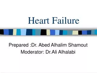
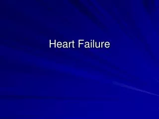
Pathophysiology • 1. Frank-Starling’s Law of the heart a. The cardiac output is a function of the preload, the afterload, and myocardial contractility. b.Preload: the volume and pressure of blood in the ventricle at the end of diastole. c. Afterload :the arterial resistance.
C 最大活动 2 正常活动 1 正常静息 心肌收缩性 左室作功 B 活动 3’ 心衰活动 3 心衰静息 静息 A D E 4 静息 致死性心肌受损 呼吸困难 肺水肿 左室舒张末容量 图3–2–1 正常和心力衰竭时对机体活动时的代偿情况
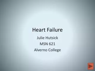
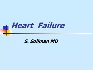
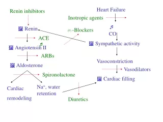
心力衰竭 + + 心肌细胞死亡 心肌细胞死亡 ↑心肌能量消耗 ↑心肌能量消耗 ↓心排血量 + - — ↑后负荷 神经体液兴奋 RAS SAS ↓心肌松弛性 ↑变力效应 心律失常 猝死 血管收缩 ↑胞浆Ca2+ InSP3 cAMP InSP3 循环 心脏 图3–2–2 肾素—血管紧张素和交感—肾 上腺素能系统激活时对心脏代偿功能的影响 2. RAAS in Heart Failure
3.myocardium impaired and remodeling initial myocardium impaired ventricular overload myocardium infarction inflammation • secondary conduct • factor • sympathetic nervous • system • RAAS • endothelins • TNF-α ,IL-6 • mechanical stress • oxidative stress disease progress heart failure complication death chamber enlargement myocardial hypertrophy embryo gene phenotype extracellular matrix change
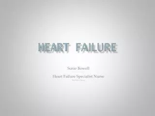
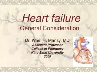
4.Diastolic heart failure • Heart failure may develop as a result of • poor ventricular filling and high filling pressure caused by abnormal ventricular relaxation
正常 容 积 压 力 顺应性↓ 顺应性↑ 图3–2–4 心室舒张末期压力和容积的关系 舒张性心力衰竭时,心室顺应性降低,心室压力–容积曲线向左上方移位,即在任何特定的舒张末期压时,心室末期容量小于正常人。
sarcoplasmic reticulum intake Ca2+ • free Ca2+ in myocyte degrade slowly • b. In CHD with obvious ischemia ,before contractility dysfunction, have occurred relaxation dysfunction • c. In hypertrophy and hypertrophic cardiomyopathy, left ventricular end-diastolic filling pressure pulmonary hypertension ,pulmonary congestion diastolic heart failure relaxation dysfunction
Type of heart failureHeart failure can be described or classified in several ways. • 1.Acute and chronic heart failure • 2.Left ,right and biventricular heart failure • 3. High and low output heart failure • 4.Diastolic and systolic dysfunction • 5.Asymptomatic and congestive heart failure
Low output heart failure: • Clinical manifestation of abnormal peripheral circulation: • vasoconstriction in system , cold, pale, extremities cyanosis, • in the late period,output per minute decrease and lead to • difference of pulse pressure decrease, the above manifestation • occur in the majority of CHF. • High output heart failure: • Extremities warm,flush, difference of pulse pressure increase, • seen in hyperthyroidism,anemia,pregnancy
Systolic dysfunction • Heart failure may develop as a result of impaired myocardial contraction . • Diastolic dysfunction • Heart failure can also be due to poor ventricular filling pressure caused by abnormal ventricular relaxation ,which is commonly found in patients with left ventricular hypertrophy, hypertension and ischemic heart disease.
§1 Chronic heart failure Definition same meaning as congestive heart failure
clinical manifestation1.left ventricular heart failuremainly manifested with pulmonary congestion and reduction of cardiac output A symptom1.dyspnea1)breathlessness2) paroxysmal nocturnal dyspnea:often with wheeze sound in both lung cardiogenic asthma
3)Orthopnea: in decubitus,blood volume flow to heart increase elevated end–diastolic filling pressure pulmonary venous and capillary pressure increase interstitial pulmonary edema pulmonary compliance decrease respiratory resistance 4)acute pulmonary edema
2. cough and hemoptysis pink-tinged or brownish sputum 3. fatigue on exertion 4. urinary system symptom in early period ,nocturia increase in later period, oliguria
B. Sign 1.general sign dyspnea after activity,also cyanosis, jaundice , difference of pulse pressure decrease, SBp decrease, rapid heart rate , peripheral vasoconstriction ,extremities cyanosis, cold, sinus tachycardia.
2.Heart sign • diffuse and laterally displaced apical impulse • gallop in early diastolic period , accentuated p2 • systolic murmur at cardiac apex • pulses alternans occur when left ventricular ejective impedance increase • 3.Lung sign • moist rales in the base of lung • ¼ CHF patients occur pleural fluid
2.Right ventricular Failure systemic circulation congestion Symptom 1)gastrointestinal tract symptom: anorexia, distention ,nausea ,vomiting ,constipation 2)kidney symptom kidney congestion renal function decrease 3)hepatic region pain: congestion , cardiac cirrhosis 4)dyspnea
Sign • 1.heart sign • heart dilate • when right heart failure is obvious,strong impulse • occur in the systolic period at the left sternal border, obvious beat occur infraxiphoid • diastolic gallop • relative tricupid incompetence • 2.hepatic cervical reflux • 3.congestive liver and tenderness occur before edema • Acute : jaundice , ALT increase • Long term: cardiac cirrhosis
4.edema occur after cervical filling and liver large, is typical sign of right heart failure. at first occur in foot, ankle , anterior tibia. In the early period,edema occur in the morning, worse in the evening ,disappear after sleeping. In the late time,systemic , symmetric, pitting edema If complicated with malnutrition or hepatic dysfunction , face edema occur, prognosis is poor. 5.pleural fluid and ascites
3.biventricular heart failure have clinical manifestation of left and right heart failure.
Conditions with normal systolic function and decreased diastolic function include: (1) systemic arterial hypertension (2) myocarditis (3) hyretrophic cardiomyopathy (4) congestive cardiomyopathy
In the setting of left ventricular dysfunction, which of following neurohormonal factors would be activated? (1)Norepinephrine (2)Endothelin (3)Arginie vasopreein (4)Endothelial-derived relaxing factor
Investigation 1.routine examination blood, urine, renal function, electrolyte, liver function 2.ECG a.no specific findings . b.Abnormalities may provide etiological clue(ventricular hypertrophy,AMI,bundle branch block) c.V1ptf<-0.03mm/s left atrial overload
3.Echocardiography:evaluating LV as well as other chamber dimensions, ejection fraction, and wall motion abnormality. a.M:obtained directing a stationary ultrasonography beam at some portion of the heart. b.Two-dimensional Echo (2-DE):provides spatially correct images of heart and has become the dominant echocardiographic modality c.Doppler Echo:using ultrasonography to record the flow of blood within the cardiovascular system.
4.X ray a evaluation of chamber enlargement b pulmonary venous congestion Kerley B lines:reflect chronic elevation of left atrial pressure and represent chronic thickening of the interlobular septa from edema. venous blood redistribution to the upper lobes. C pulmonary venous pressure>25-30mmHg(3.3-4KPa) interstitial edema occur.
参 数 正常值 临床意义 中心静脉压(CVP) 6~12cmH2O(0.59~1.18KPa) ↑说明血容量过多或右心衰竭 肺动脉压(PAP) 12~30/4~13mmHg(1.6~4.0/0.53~1.73KPa) ↑说明肺动脉高压、左心衰竭 肺毛细血管楔嵌压(PCWP) 6~12mmHg(0.8~1.6KPa) ↑说明肺淤血、左心衰竭 心搏量(SV) 60~70ml ↓可由于前负荷不足、心包填塞、 心肌收缩力下降,心排阻力上升 心搏指数(SI) 41~51ml/m2 同上 心排血量(CO) 5~6L/min ↑可由于正性肌力药物作用, ↓说明有心力衰竭 心排指数(CI) 2.6~4.0L/(min·m2) ↓说明收缩力减低或心力衰竭 射血分数(EF) 0.5~0.6 ↓说明心室收缩功能减低 左室每搏作功(LVSW) 60~123 左室每搏作功指数(LVSWI) 50~62 体循环血管阻力(SVR) 770~1500dynes·s/ cm5 ↓见于缺血、血管扩张剂, ↑高血压、血管活性药物 体循环血管阻力指数(SVRI) 1970~2390dynes·s(cm5·m2) 同上 肺血管阻力(PVR) 37~250 dynes·s/ cm5 ↑毛细血管前肺小动脉收缩、肺栓塞、慢性肺疾病、肺间质水肿、肺小血管阻塞性病变、二尖瓣狭窄 肺血管阻力指数(PVRI) 69~177 dynes·s(cm5·m2) 同上 ↑增高 ↓降低 Invasive homodynamic monitoring
Diagnosis and differential diagnosis Clinical diagnosis include : etiology(basic cause and induce cause), pathoanatomy, pathophysiology, heart rhythm cardiac function
NYHA classification Ⅰ no activity limit , daily activity don't lead to inertia, dyspnea, palpitation. Ⅱ slight activity limit , no symptom at rest ,daily activity lead to inertia, dyspnea, palpitation or angina pectoris. Ⅲ obvious activity limit , no symptom at rest , daily activity lead to inertia, dyspnea, palpitation or angina pectoris. Ⅳ cannot do any activity , have symptom at rest.
type CI (L/min·m2) PCWP (mmHg) Clinical manifestation Ⅰ ≥2.2 ≤18(2.4) No peripheral perfusion deficiency and pulmonary congestion ,no symptom and sign of heart failure Ⅱ ≥2.2 >18(2.4) No peripheral perfusion deficiency ,pulmonary congestion ,no obvious clinical manifestation Ⅲ <2.2 ≤18(2.4) peripheral perfusion deficiency, no pulmonary congestion ,seen in right ventricular infarction and blood volume deficiency Ⅳ <2.2 >18(2.4) peripheral perfusion deficiency and pulmonary congestion ,severe type Forrester classification
Killip classification Ⅰ no heart failure symptom, no moist rales, PCWP may elevate Ⅱ slight to moderate heart failure, <50% lung field moist rales, S3 gallop, persist sinus tachycardia ,x ray manifestation of pulmonary congestion Ⅲ severe heart failure, >50%lung field moist rales, may occur lung edema Ⅳ cardiac shock, Bp<90mmHg, oliguria <20ml/h, skin cold, cyanosis, tachypnea, rapid pulse V cardiogenic shock and pulmonary edema
Differential diagnosis 1. Left heart failure Pulmonary ,cardiogenic dyspnea 2. Right heart failure constrictive pericarditis renal edema hepatic cirrhosis
Management of heart failure 1.Etiologic treatment basic cause , precipitating causes. 2.Reduction of ventricular overload a.rest and sedative agent b.salt-intake control normal adult intake 3-6g salt per day Ⅰ0 heart failure : 2g salt/per day Ⅱ 0heart failure : 1g salt/per day Ⅲ 0heart failure : 0.4g salt/per day
c.water intake control may not limit water intake strictly, intake water 1.5-2.0L per day in severe heart failure, water retention, seral albumin decrease, dilutive hyponatremia, not only limit salt intake ,but also control water intake
利尿剂 作用部位和机制 剂量(mg/d) 作用持续时间(h) d.diuretics 排钾类 氢氯噻嗪 (hydrochlorothiozide) 远曲小管:抑制NaCl共转运 25~100口服 12~18 美托拉宗 (metolazone) 同上 5~20口服 12~24 氯噻酮 (chlorothalidone 同上 25~100口服 24~72 呋噻米 (furosemide) Henle襻上升支:抑制Na-K-2Cl转运 20~1000口服/静注 4~6 丁脲酸 (bumetanide) 同上 0.5~20口服 4~6 潴钾类 氨体舒通 (spironolactone 集合管:醛固酮拮抗剂 25~100口服 24~96 氨苯喋啶 (triamterene) 集合管:抑制Na重吸收 100~300口服 12~16 阿米洛利 (amiloride) 同上 5~20口服 12~18
Reasonable application of diuretics 1. strictly following indication 2. combined medication K-sparing diuretics is contradicted in renal dysfunction 3. intermissional therapy 4. Pay attention to water and electrolyte disturbance
Differential of deficit sodium and diluted hyponatremia deficit sodium hyponatremia occurred after using many diuretics. feature: postohypotension, oliguria, high urine gravity, should intake salt diluted hyponatremia also called refractory heart failure ,hyponatremia of high blood volume ,should limit water-intake
e.Vasodilator drugs Indication 1.Left end-diastolic filling pressure>18mmHg, pulmonary congestion 2.clinical manifestation of peripheral circulatory perfusion deficiency CI<2.2L/min.m2 3.valve insufficiency, ventricular septal defect pulmonary hypertension,valve regurgitation with cardiac dysfunction If blood volume deficiency ,should fluid replacement at first,then use vasodilator drugs.
药物 机制 前负荷 后负荷 常用剂量 作用时间 开始 高峰 持续 硝酸盐血管扩张剂 硝酸甘油 NO供者 +++ + 0.2~10μg/(kg·min) iv 5~6mg 经皮 2min 5~15min <30min 二硝酸异山梨醇酯 +++ + 10~60mg po tid 15~20min 1h 4h 10~20mg 舌下 5min 15~30min 3h 2~7mg/h iv 3~5min 2h 3h 硝普钠 +++ +++ 0.1~0.3μg/(kg·min) iv 几乎立即 停药2~15min消失 交感神经阻滞剂 酚妥拉明 非选择性α–肾上腺素能激动剂 ++ ++ 0.5~1.0mg/min iv 15~20min 3~4h 哌唑嗪 α1–肾上腺素能受体拮抗剂 +++ ++ 1~6mg po tid 30min 1~3h 6h 肾素–血管紧张素系统拮抗剂 卡托普利 抑制由ACE引起的肾系统性生 成和组织生成血管紧张素Ⅱ; 降低缓激肽的代谢 ++ ++ 6.25~50mg po q8h 15~30min 1~2h 4~6h 依那普利 ++ ++ 5~10mg po bid 2h 4~6h 24h 赖诺普利 ++ ++ 2.5~20mg po q12~24h 6~8h 12h 雷米普利 ++ ++ 1.25~5mg po qd 1~2h 3~6h 24h 芦沙坦 阻断血管紧张素Ⅱ(AT1受体) ++ ++ 25~50mg po q12h 5~6h 24h