Download

1 / 82
820 likes | 882 Vues
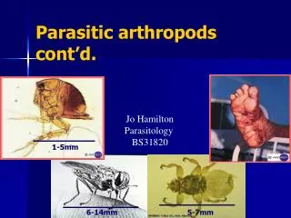
A research on 45,128 individuals showed a 8.45% male and 61% urban residents infection rate. Parasitic infection was prevalent among all age groups. Recommendations for reducing the issue are highlighted in the findings.

E N D
از مجموع 53995 نفر نمونه مورد بررسی، تحقيق روی 45128 نفر که 8/45% جنس مذکر و 61% مربوط به ساکنين مناطق شهری بودند، انجام گرفت. • آلودگی در3 /19% وجود داشت که در مردان7 /19 و در زنان1 /19 درصد بود. • در بين انواع تک ياخته، ژيارديا با9 /10% و از بين کرم ها آسکاريس با5 /1% از شيوع بيشتر برخوردار بودند. • آلودگی در گروه سنی کمتر از 15 سال به ميزان5 /25% و در سنين بالای 70 سال به ميزان6 /11 بود. • ميزان آلودگی در ساکنين شهری4 /16% و در روستاييان3 /24% گزارش شد. • نتيجه گيری و توصيه ها: ميزان عفونت انگلی روده ای در ايران جای نگرانی دارد. با توجه به عوارض شناخته شده، بررسی علل آن و اجرا برنامه ها برای کاهش مشکل توصيه می شود.
Parasites are divided into 2 main groups taxonomically: protozoans, which are unicellular, and helminths, which are multicellular. • Chemotherapeutic agents appropriate for 1 group may not be appropriate for the other, and not all drugs are readily available.
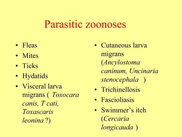
Protozoan Diseases • Primary Amebic Meningoencephalitis • Amebiasis • Giardiasis and Balantidiasis • Cryptosporidium, Isospora, Cyclospora, and Microsporidia • Trichomoniasis (Trichomonas vaginalis) • Leishmaniasis(Leishmania) • African Trypanosomiasis (Sleeping Sickness; Trypanosoma brucei Complex) • American Trypanosomiasis (Chagas Disease; Trypanosoma cruzi) • Malaria(Plasmodium) • Babesiosis (Babesia) • Toxoplasmosis(Toxoplasma gondii)
Amebiasis • Entamoeba histolytica infects up to 10% of the world's population; endemic foci are particularly common in the tropics, especially in areas with low socioeconomic and sanitary standards. • In most infected individuals, E. histolytica parasitizes the lumen of the gastrointestinal tract and causes few symptoms or sequelae. • The 2 most common forms of disease caused by E. histolytica are amebic colitis with parasitic invasion of the intestinal mucosa and amebic liver abscesswith dissemination of the parasite to the liver.
Etiology • E. histolytica, the pathogenic species, causes a spectrum of disease and can become invasive. • Infection is acquired through the ingestion of parasite cysts, which measure 10-18 mm in diameter and contain 4 nuclei. • Cysts are resistant to harsh environmental conditions including the concentrations of chlorine commonly used in water purification but can be killed by heating to 55C.
After ingestion, cysts are resistant to gastric acidity and digestive enzymes and germinate in the small intestine to form trophozoites. • These large, actively motile organisms colonize the lumen of the large intestine and may invade the mucosal lining. • Infection is not transmitted by trophozoites, as these rapidly degenerate outside the body and are unable to survive the low pH of the stomach if swallowed.
Epidemiology • It is estimated that infection with E. histolytica leads to 50 million cases of symptomatic disease and 40,000-110,000 deaths annually. • Prospective studies have shown that 4-10% of individuals infected with E. histolytica develop amebic colitis and that <1% of infected individuals develop disseminated disease, including amebic liver abscess.
Clinical Manifestations • Clinical presentations range from asymptomatic cyst passage to amebic colitis, amebic dysentery, ameboma, and extraintestinal disease. • E. histolytica infection is asymptomatic in about 90% of persons but has the potential to become invasive and should be treated. • Severe disease is more common in young children, pregnant women, malnourished individuals, and persons taking corticosteroids. • Extraintestinal disease usually involves the liver, but less common extraintestinal manifestations include amebic brain abscess, pleuropulmonary disease, ulcerative skin, and genitourinary lesions.
Diagnosis • A diagnosis of amebic colitis is made in the presence of compatible symptoms with detection of E. histolytica antigens in stool. • This approach has a greater than 95% sensitivity and specificity and coupled with a positive serology test is the most accurate means of diagnosis in developed countries. • The E. histolytica II stool antigen detection test (TechLab, Blacksburg, VA) is able to distinguish E. histolytica from E. dispar infection. • Microscopic examination of stool samples has a sensitivity of 60%. • Sensitivity can be increased to 85-95% by examining 3 stools, since excretion of cysts can be intermittent. • However, microscopy cannot differentiate between E. histolytica and E. dispar unless phagocytosed erythrocytes (specific for E. histolytica) are seen.
Differential diagnosis • The differential diagnosis for amebic colitis includes colitis due to bacterial (Shigella, Salmonella, enteropathogenic Escherichia coli, Campylobacter, Yersinia, Clostridium difficile), mycobacterial (tuberculosis and atypical mycobacteria), and viral (cytomegalovirus) pathogens, as well as noninfectious causes such as inflammatory bowel disease (IBD). • Pyogenic liver abscess due to bacterial infection, hepatoma, and echinococcal cysts are in the differential for amebic liver abscess. • However, echinococcal cysts are rarely associated with systemic symptoms such as fever unless there is cyst rupture or leakage.
Treatment • Invasive amebiasis is treated with a nitroimidazole such as metronidazole or tinidazole and then a luminal amebicide.
Prevention • Control of amebiasis can be achieved by exercising proper sanitation and avoiding fecal-oral transmission. • Regular examination of food handlers and thorough investigation of diarrheal episodes may help identify the source of infection. • No prophylactic drug or vaccine is currently available for amebiasis.
Giardiasis • Giardia lambliais a flagellated protozoan that infects the duodenum and small intestine. • Infection results in clinical manifestations that range from asymptomatic colonization to acute or chronic diarrhea and malabsorption. • Infection is more prevalent in children than in adults.
Giardia is endemic in areas of the world with poor levels of sanitation. • It is also an important cause of morbidity in developed countries, where it is associated with urban child-care centers, residential institutions for the developmentally delayed, and water-borne and food-borne outbreaks. • Giardia is a particularly significant pathogen in people with malnutrition, certain immunodeficiencies, and cystic fibrosis.
Etiology • The life cycle of G. lamblia (also known as Giardia intestinalis or Giardia duodenalis) is composed of 2 stages: trophozoites and cysts. • Giardia infects humans after ingestion of as few as 10-100 cysts (which measure 8-10 mm in diameter). • Each ingested cyst produces 2 trophozoites in the duodenum. • After excystation, trophozoites colonize the lumen of the duodenum and proximal jejunum, where they attach to the brush border of the intestinal epithelial cells and multiply by binary fission.
Epidemiology • Giardia occurs worldwide and is the most common intestinal parasite identified in public health laboratories in the USA, where it is estimated that up to 2 million cases of giardiasis occur annually. • The age-specific prevalence of giardiasis is high during childhood and begins to decline after adolescence. • The asymptomatic carrier rate of G. lamblia in the USA is as high as 20-30% in children younger than 36 mo of age attending child-care centers. • Asymptomatic carriage may persist for several months.
Transmission of Giardia is common in certain high-risk groups, including children and employees in child-care centers, consumers of contaminated water, travelers to certain areas of the world, men who have sex with men, and persons exposed to certain animals. • Humoral immunodeficiencies, including common variable hypogammaglobulinemia and X-linked agammaglobulinemia, predispose humans to chronic symptomatic Giardia infection, suggesting the importance of humoral immunity in controlling giardiasis. • Selective immunoglobulin A (IgA) deficiency is also associated with Giardia infection.
Clinical Manifestations • The incubation period of Giardia infection usually is 1-2 wk but may be longer. • A broad spectrum of clinical manifestations occurs, depending on the interaction between G. lamblia and the host. • Children who are exposed to G. lamblia may experience asymptomatic excretion of the organism, acute infectious diarrhea, or chronic diarrhea with persistent gastrointestinal tract signs and symptoms, including failure to thrive and abdominal pain or cramping.
Symptomatic infections occur more frequently in children than in adults. • Most symptomatic patients usually have a limited period of acute diarrheal disease with or without low-grade fever, nausea, and anorexia; in a small proportion of patients, an intermittent or more protracted course characterized by diarrhea, abdominal distention and cramps, bloating, malaise, flatulence, nausea, anorexia, and weight loss develops.
Diagnosis • Giardiasis should be considered in children who have acute nondysenteric diarrhea, persistent diarrhea, intermittent diarrhea and constipation, malabsorption, chronic crampy abdominal pain and bloating, failure to thrive, or weight loss. • It should be particularly high in the differential diagnosis of children in child care, children in contact with an index case, children with a history of recent travel to an endemic area, and children with humoral immunodeficiencies. • Testing for giardiasis should be standard for internationally adopted children from Giardia-endemic areas, and screening for iron deficiency should be considered in internationally adopted children with giardiasis.
Stool enzyme immunoassay (EIA) or direct fluorescent antibody tests for Giardia antigens are now the tests of choice for giardiasis in most situations. EIA is less reader dependent and more sensitive for detection of Giardia than microscopy. • Traditionally, a diagnosis of giardiasis has been established by microscopy documentation of trophozoites or cysts in stool specimens, but 3 stool specimens are required to achieve a sensitivity of >90%. • In patients with chronic symptoms in whom giardiasis is suspected but in whom testing of stool specimens for Giardia yields a negative result, aspiration or biopsy of the duodenum or upper jejunum should be considered. • In a fresh specimen, trophozoites usually can be visualized by direct wet mount.
Treatment • Children with acute diarrhea in whom Giardia organisms are identified should receive therapy. • In addition, children who manifest failure to thrive or exhibit malabsorption or gastrointestinal tract symptoms such as chronic diarrhea should be treated. • Asymptomatic excreters generally are not treated except in specific instances such as in outbreak control, for prevention of household transmission by toddlers to pregnant women and patients with hypogammaglobulinemia or cystic fibrosis, and in situations requiring oral antibiotic treatment where Giardia may have produced malabsorption of the antibiotic.
Prevention • Infected persons and persons at risk should practice strict handwashing after any contact with feces. • This point is especially important for caregivers of diapered infants in child-care centers, where diarrhea is common and Giardia organism carriage rates are high.
Leishmaniasis (Leishmania) • The leishmaniases are a diverse group of diseases caused by intracellular protozoan parasites of the genus Leishmania, which are transmitted by phlebotomine sandflies. • Multiple species of Leishmania are known to cause human disease involving the skin and mucosal surfaces and the visceral reticuloendothelial organs. • Cutaneous disease is generally mild but may cause cosmetic disfigurement. • Mucosal and visceral leishmaniasis is associated with significant morbidity and mortality.
Visceral Leishmaniasis • VL (kala-azar) typically affects children <5 yr of age in the New World and Mediterranean region (L. infantum/chagasi) and older children and young adults in Africa and Asia (L. donovani). • After inoculation of the organism into the skin by the sandfly, the child may have a completely asymptomatic infection or an oligosymptomatic illness that either resolves spontaneously or evolves into active kala-azar.
Children with asymptomatic infection are transiently seropositive but show no clinical evidence of disease.
Children who are oligosymptomatic have mild constitutional symptoms (malaise, intermittent diarrhea, poor activity tolerance) and intermittent fever; most will have a mildly enlarged liver. • In most of these children the illness will resolve without therapy, but in approximately 25% it will evolve to active kala-azar within 2-8 mo. • Extreme incubation periods of several years have rarely been described. • During the 1st few weeks to months of disease evolution the fever is intermittent, there is weakness and loss of energy, and the spleen begins to enlarge.
The classic clinical features of high fever, marked splenomegaly, hepatomegaly, and severe cachexia typically develop approximately 6 mo after the onset of the illness, but a rapid clinical course over 1 mo has been noted in up to 20% of patients in some series. • At the terminal stages of kala-azar the hepatosplenomegaly is massive, there is gross wasting, the pancytopenia is profound, and jaundice, edema, and ascites may be present. • Anemia may be severe enough to precipitate heart failure. • Bleeding episodes, especially epistaxis, are frequent. • The late stage of the illness is often complicated by secondary bacterial infections, which frequently are a cause of death.
A younger age at the time of infection and underlying malnutrition may be risk factors for the development and more rapid evolution of active VL. • Death occurs in >90% of patients without specific antileishmanial treatment.
Laboratory Findings • Laboratory findings associated with classic kala-azar include anemia (hemoglobin 5-8 mg/dL), thrombocytopenia, leukopenia (2,000-3,000 cells/micL), elevated hepatic transaminase levels, and hyperglobulinemia (>5 g/dL) that is mostly immunoglobulin G (IgG).
Differential Diagnosis • VL should be strongly suspected in the patient with prolonged fever, weakness, cachexia, marked splenomegaly, hepatomegaly, cytopenias, and hypergammaglobulinemia who has had potential exposure in an endemic area. • The clinical picture may also be consistent with that of malaria, typhoid fever, miliary tuberculosis, schistosomiasis, brucellosis, amebic liver abscess, infectious mononucleosis, lymphoma, and leukemia.
Diagnosis • Serologic testing by enzyme immunoassay, indirect fluorescence assay, or direct agglutination is very useful in VL because of the very high level of antileishmanial antibodies. • An enzyme-linked immunosorbent assay using a recombinant antigen (K39) has a sensitivity and specificity for VL that is close to 100%. • A negative serologic test result in an immunocompetent individual is strong evidence against a diagnosis of VL. • Serodiagnostic tests have positive findings in only about half of the patients who are co-infected with HIV.
In patients with VL, smears or cultures of material from splenic, bone marrow, or lymph node aspirations are usually diagnostic. • In experienced hands, splenic aspiration has a higher diagnostic sensitivity, but it is rarely performed in the USA because of the risk for bleeding complications.
Treatment • All patients with VL or ML should receive therapy. • Glucantime • Amphotericin B
Malaria (Plasmodium) • Malaria is an acute and chronic illness characterized by paroxysms of fever, chills, sweats, fatigue, anemia, and splenomegaly. • It has played a major role in human history, causing harm to more people than perhaps any other infectious disease. • Malaria is of overwhelming importance in the developing world today, with an estimated 300 to 500 million cases and more than 1 million deaths each year. • Most malarial deaths occur among infants and young children.
Etiology • Malaria is caused by intracellular Plasmodium protozoa transmitted to humans by female Anopheles mosquitoes. • Prior to 2004, only 4 species of Plasmodium were known to cause malaria in humans: P. falciparum, P. malariae, P. ovale, and P. vivax. • In 2004 P. knowlesi (a primate malaria species) was also shown to cause human malaria, and cases of P. knowlesi infection have been documented in Malaysia, Indonesia, Singapore, and the Philippines.
Malaria also can be transmitted through blood transfusion, use of contaminated needles, and from a pregnant woman to her fetus. • The risk for blood transmission is small and decreasing in the USA but may occur by way of whole blood, packed red blood cells, platelets, leukocytes, and organ transplantation.
Clinical Manifestations • The classic presentation of malaria is seldom noted with other infectious diseases and consists of paroxysms of fever alternating with periods of fatigue but otherwise relative wellness. • Febrile paroxysms are characterized by high fever, sweats, and headache, as well as myalgia, back pain, abdominal pain, nausea, vomiting, diarrhea, pallor, and jaundice.
Typical laboratory findings include anemia, thrombocytopenia, and a normal or low leukocyte count. The erythrocyte sedimentation rate (ESR) is often elevated.
WORLD HEALTH ORGANIZATION CRITERIA FOR SEVERE MALARIA, 2000 Impaired consciousness Prostration Respiratory distress Multiple seizures Jaundice Hemoglobinuria Abnormal bleeding Severe anemia Circulatory collapse Pulmonary edema
Diagnosis • Any child who presents with fever or unexplained systemic illness and has traveled or resided in a malaria-endemic area within the previous year should be assumed to have life-threatening malaria until proven otherwise. • Malaria should be considered regardless of the use of chemoprophylaxis. • Important criteria that suggest P. falciparum malaria include symptoms occurring less than 1 mo after return from an endemic area, more than 2% parasitemia, ring forms with double chromatin dots, and erythrocytes infected with more than 1 parasite.
The diagnosis of malaria is established by identification of organisms on Giemsa-stained smears of peripheral blood or by rapid immunochromatographic assay.
Differential Diagnosis • The differential diagnosis of malaria is broad and includes viral infections such as influenza and hepatitis, sepsis, pneumonia, meningitis, encephalitis, endocarditis, gastroenteritis, pyelonephritis, babesiosis, brucellosis, leptospirosis, tuberculosis, relapsing fever, typhoid fever, yellow fever, amebic liver abscess, Hodgkin disease, and collagen vascular disease.
Treatment • Fever without an obvious cause in any patient who has left a P. falciparum endemic area within 30 days and is nonimmune should be considered a medical emergency. • Thick and thin blood smears should be obtained immediately, and all children with symptoms of severe disease should be hospitalized. • If blood films are negative, they should be repeated every few hours.