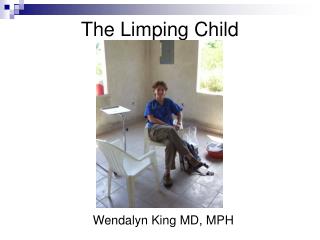
THE LIMPING CHILD
290 likes | 771 Vues
THE LIMPING CHILD. PRESENTED BY DANIEL L. MORRISON, D.O. CLINICAL PROFESSOR, MICHIGAN STATE UNIVERSITY COLLEGE OF OSTEOPATHIC MEDICINE. Introduction.

THE LIMPING CHILD
E N D
Presentation Transcript
THE LIMPING CHILD PRESENTED BY DANIEL L. MORRISON, D.O. CLINICAL PROFESSOR, MICHIGAN STATE UNIVERSITY COLLEGE OF OSTEOPATHIC MEDICINE
Introduction • Limping is a common problem in children and adolescents. The different diagnoses of limping is extensive and includes numerous abnormalities of the lower extremity and spine.
Common conditions that can cause a child to limp: • Conditions divided into two categories: • Antalgic • Trendelenburg
Antalgic definition • Painful limp • The child spends the greater portion of the gait cycle on the asymptomatic leg than the symptomatic.
Antalgic • Infectious • Septic arthritis • Osteomyelitis • Acute • Subacute • Diskitis • Rheumatologic • Juvenile arthritis
Antalgiccont. • Trauma • Sprains,strains, contusions • Fractures • Toddler’s fx • Stress fx • Be aware of child abuse
Antalgiccont. • Neoplasia • Benign • Osteoid osteoma • Malignant • Osteogenic sarcoma • Ewing sarcoma • Leukemia • Spinal cord tumors
Antalgiccont. • Congenital • Tarsal coalition • Acquired • Legg-Calve-Perthes disease • Slipped capital femoral epiphysis
Trendelenburg • Dr. Friedrich Trendelenberg born in Berlin in 1844. • Classic article reproduced the gait of patients with congenital dislocations of the hip.
Trendelenburg’s Sign • Positive sign shows the pelvis hanging down on the swinging side • Negative sign show the pelvis angled up on the swinging side
Trendelenburg Limp • Developmental dysplasia of the hip • Leg length discrepancy • Neuromuscular Disease • Cerebral palsy • Muscular dystrophy
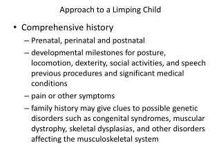
Clinical History • Begin with thorough history, family history, and physical examination • Onset (acute-insidious) • Age (chronological-developmental) • Symptom complex
Differential Diagnosis based upon age • Toddler (1-3 years of age) • Childhood (4-10 years) • Adolescence (11+ years)
Differential for Toddlers • Infection • Septic arthritis-hip,knee • Osteomyelitis • Diskitis
Differential for Toddlers cont. • Occult trauma • Sprains, strains, contusions • Toddler’s fx • Stress fx
Differential for Childhood • Infection • Septic arthritis of hip or knee • Osteomyelitis • Diskitis
Differential for Childhoodcont. • Transient synovitis of the hip • Legg-Calve-Perthes disease • Juvenile arthritis • Trauma • Neoplasia • Leg length discrepancy
Differential for Adolescence • Slipped capital femoral epiphysis • Juvenile arthritis • Trauma • Leg length discrepancy
Differential for Adolescence cont. • Neoplasia • HNP • Congenital Spine • Spina Bifida Occulta • Spondylolisthesis • L5 radiculopathy
Physical Examination • Observing the child’s walk after removing all clothing except diaper or underwear and having the child walk a sufficient distance to observe the gait pattern.
Gait Analysis • Stance Phase • Heel strike, foot flat, midstance, heel off, toe off • Swing Phase • Acceleration, mid swing, deceleration
Distinguishing characteristics: • redness, swelling, tenderness • abrasion suggesting trauma • café au lait spots • rash
Characteristics cont. • joint effusions • soft tissue masses • alteration of strength, sensation, or DTRs
Laboratory Assessment • Blood cultures • WBC count with differential • Erythrocyte sedimentation rate • C-reactive protein level • Antinuclear antibody
Imaging Modalities • Plain Radiographs • Bone Scan • Ultrasound • Computed Topography • MRI