Download
1 / 33
330 likes | 375 Vues
Learn about bone fractures, complications like fat embolism and compartment syndrome, and treatments such as bone grafting, stimulation methods, and surgeries in orthopedic trauma care.

E N D
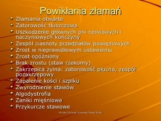
Powikłania złamań • Złamania otwarte • Zatorowość tłuszczowa • Uszkodzenie głównych pni nerwowych i naczyniowych kończyny • Zespół ciasnoty przedziałów powięziowych • Zrost w nieprawidłowym ustawieniu • Zrost opóźniony • Brak zrostu (staw rzekomy) • Zakrzepica żylna: zatorowość płucna, zespół pozakrzepowy • Zapalenie kości i szpiku • Zwyrodnienie stawów • Algodystrofia • Zaniki mięśniowe • Przykurcze stawowe Klinika Chirurgii Urazowej Paweł Grala
Stimulation of bone healing • Osteognesis – formation of new bone by existing differentiated bone forming cells of any origin • Osteoinduction – differentiation of previously uncommitted conective tissue cells into bone forming cells • Osteoconduction – inert material acts as a scaffold for bone formation by gradual substitution (ingrowth of cells and blood vessels). Bone grafting, mechanical stimulation, electical stimulation, ultrasound, vascular stimulation, biological stimulation Klinika Chirurgii Urazowej Paweł Grala
Compartment syndrome Klinika Chirurgii Urazowej Paweł Grala
Compartment syndrome • Increased pressure within a limited space (unyielding fascia) compromises the circulation and function of the tissues contained within that space • Results in ischaemic muscle damage nerve damage, renal failure, infection, amputation, or even death. • Damaged muscle heals by fibrosis, causing shortening of the musculo-tendinous unit (joint stiffness, cavus foot with clawing of the toes and claw hand deformity) • Deformities appear within the first year after injury. Klinika Chirurgii Urazowej Paweł Grala
Fasciotomy Klinika Chirurgii Urazowej Paweł Grala
Compartment syndrome Klinika Chirurgii Urazowej Paweł Grala
Compartment syndrome Klinika Chirurgii Urazowej Paweł Grala
Fat embolism – presence of fat globules in the lung parenchyma and peripheral circulation after a long bone fracture or major trauma (90% of patients with long bone fractures, inj. to fat tissue). Fat embolism syndrome (1%) – clinical manifestation of FE (within 72 h of injury): -progressive respiratory insufficiency (hypoxia) -thrombocytopenia (<150th) -deteriorating mental condition -tachycardia (>120) -petechiae -pyrexia (>39°C) -hematocrit decrease -many confounding physiological derangements -supportive treatment Klinika Chirurgii Urazowej Paweł Grala
100 patients undergoing IM nailing of the femur: FES – 11% in delayed fixation group 0% in early fixation group Klinika Chirurgii Urazowej Paweł Grala
DVT • It is estimated that 100,000 patients in the United States die from PE each year. Pulmonary embolism has been observed in 2%-22% of patients with trauma, and fatal PE is the third most common cause of death in trauma patients who survive the first 24 hours. • Virchow elucidated the pathogenesis of VTE in 1856. Substantial evidence exists demonstrating that trauma survivors experience significant venostasis, venous valvular and intimal injury, and periods of intense hypercoagulability. Some evidence suggests that DVT in trauma develops against a background of activated coagulation. • hypercoagulability persist for at least one month after injury in 80% of patients. • Most thrombi in high-risk patients begin in the deep veins of the calf, do not extend proximally, remain asymptomatic,12,13 and it has been estimated that 10%-20% of calf thrombi extend into the proximal veins.14,15 From these proximal thrombi, 50% eventually lead to PE and 10% of patients with PE die of this complication Klinika Chirurgii Urazowej Paweł Grala
„life over limb” principle Klinika Chirurgii Urazowej Paweł Grala
Malunion • A malunited fracture is one that has healed with the fragments in a nonanatomic position. • May impair function in several ways: (1) an abnormal joint surface may cause irregular weight transfer and arthritis of the joint, especially in the lower extremities; (2) rotation or angulation of the fragments may interfere with proper balance or gait in the lower extremities or positioning of the upper extremities; (3) overriding of fragments or bone loss may result in perceptible shortening; and (4) the movements of neighboring joints may be blocked. • Are commonly the rule in the closed treatment of fractures; • A malunited fracture becomes surgically significant only when it impairs function. Klinika Chirurgii Urazowej Paweł Grala
Malunion • Characteristics that determine the acceptability of fracture reduction: 1. alignment 2. rotation 3. restoration of normal length 4. actual position of the fragments (least important). A slight deformity may be seriously disabling when a malunion involves a joint, is more important in lower extraemities and small bones Klinika Chirurgii Urazowej Paweł Grala
Nonunion • Union is considered delayed when healing has not advanced (clinical and radiographic) at the average rate for the location and type of fracture. • FDA definiton „n. is established when a minimum of 9 months has elapsed since injury and the fracture shows no visible progresive signs of healing for 3 months.„ • Process of scar formation in which the rate of endosteal and periosteal osteogenesis is low, being outweighed by bone resorption • The final status of a nonunited fracture is the formation of a pseudarthrosis Klinika Chirurgii Urazowej Paweł Grala
nonunion • Often in fractures that were: (1) open, (2) infected, (3) segmental, with impaired blood supply, usually to the middle fragment, (4) comminuted by severe trauma, (5) insecurely fixed, (6) immobilized for an insufficient time, (7) treated by ill-advised open reduction, or (8) distracted either by traction or by a plate and screws (9) long non-weigt bearing • Most common in the tibia (2%) • v.important – quality of soft tissue coverage, mobility of bone fragments. • Treatment: realignement, stabilization, stimulation Klinika Chirurgii Urazowej Paweł Grala
Avascular nonunions (atrophic,inert) - incapable of biologic reaction. • Torsion wedge nonunions - presence of an intermediate fragment in which the blood supply is decreased or absent. The fragment has healed to one main fragment but not to the other (typically seen in tibial fractures treated by plate and screws). • Comminuted nonunions - presence of one or more intermediate fragments that are necrotic. The roentgenograms show absence of any sign of callus formation. Typically these nonunions result in the breakage of any plate used in stabilizing the acute fracture. • Defect nonunions - loss of a fragment of the diaphysis of a bone. The ends of the fragments are viable, but union across the defect is impossible. As time passes the ends of the fragments become atrophic (after open fractures, sequestration in osteomyelitis and resection of tumors). • Atrophic nonunions - final result when intermediate fragments are missing and scar tissue that lacks osteogenic potential is left in their place. The ends of the fragments have become osteoporotic and atrophic. Klinika Chirurgii Urazowej Paweł Grala
Atrophic nonunion Klinika Chirurgii Urazowej Paweł Grala
Hypervascular nonunion (hypertrophic, viable) - capable of biologic reaction. • "Elephant foot" – hypertrophic, rich in callus. Result from insecure fixation or premature weight bearing in a reduced fracture whose fragments are viable. • „Horse hoof" mildly hypertrophic and poor in callus. Typically occur after a moderately unstable fixation with plate and screws. The ends of the fragments show some callus, insufficient for union, and possibly a little sclerosis. • Oligotrophic - not hypertrophic, callus is absent. They typically occur after major displacement of a fracture, distraction of the fragments, or internal fixation without accurate apposition of the fragments. Klinika Chirurgii Urazowej Paweł Grala
Hypervascular nonunion (hypertrophic, viable)Elephant foot Horse hoof Oligotrophic Klinika Chirurgii Urazowej Paweł Grala
Femoral nonunion Klinika Chirurgii Urazowej Paweł Grala
Ulnar pseudoarthrosis Klinika Chirurgii Urazowej Paweł Grala
Kryteria diagnostycznealgodystrofii • Objawy kliniczne: 1.ból 2.obrzęk 3.zmiana zabarwienia 4.ucieplenia i 5.potliwości skóry, 6.zesztywnienie stawów. • Rozpoznanie gdy co najmniej 4 objawy w okolicy nie związanej bezpośrednio z urazem. • Dodatkowo zmiany troficzne paznokci i włosów. Klinika Chirurgii Urazowej Paweł Grala
Leczenie algodystrofiipodział wg. Steinbrockera • W I fazie (hypertroficzna-ciepła) Calcihexal s.c. co 2 dni 100g/1-2miesiące, Diclofenac 2 x 0,05 • w II fazie (hypotroficzna-zimna) Diclofenac j.w. i Polpressin 2 x 0,005, oraz Provasan 3 x 0,05. Klinika Chirurgii Urazowej Paweł Grala
Problemy • Diagnostyka - objawy kliniczne\scyntygrafia kostna • Diagnostyka - scyntygrafia naczyniowa (etapy choroby, leczenie?) • Wymiatacze wolnych rodników tlenowych np. Vit.C 500mg/d (teorie etiologiczne) • czynniki ryzyka (stopień uszkodzenia kości, ucisk nerwu pośrodkowego) Klinika Chirurgii Urazowej Paweł Grala
C-spine radiographs • Low risk: no neck pain or tenderness, no neurologic signs, no history of loss of consciousness, normal mental status, no distracting injury • Other – collar in place, L with C7-T1 junction (swimmers view), AP, open mouth odontoid view, flexion-extension views in cooperating patients (no passive motion) • CT – limited ability to show ligamentous injuries Klinika Chirurgii Urazowej Paweł Grala