ASCARIASIS
380 likes | 1.01k Vues
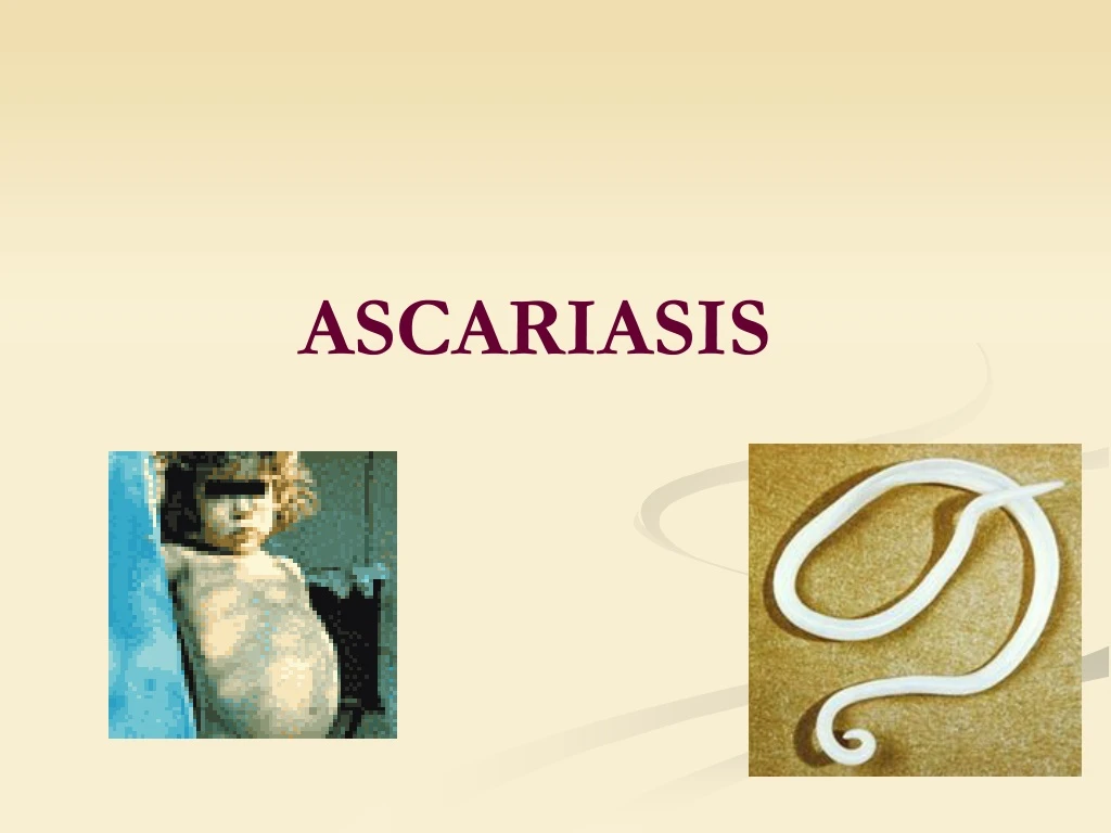
ASCARIASIS . Ascariasis – is a intestinal parasitic disease caused by species of round worm of the class Nematoda, and can cause intestinal and lung damage. Ascaris lumbricoides, an intestinal roundworm, is one of the most common helminthic human infections worldwide.

ASCARIASIS
E N D
Presentation Transcript
Ascariasis – is a intestinal parasitic disease caused by species of roundworm of the class Nematoda, and can cause intestinal and lung damage. • Ascaris lumbricoides, an intestinal roundworm, is one of the most common helminthic human infections worldwide. • Highest prevalence in tropical and subtropical regions, and areas with inadequate sanitation. Ascariasis occurs in rural areas of the southeastern United States.
Morphology • A. lumbricoides is the largest intestinal nematode of man. • The female worms are larger than the males and can measure 40 cm in length and 6 mm in diameter. • They are white or pink and are tapered at both ends. • The ova are oval, have a thick shell, a mamillated outer coat.
EPIDEMIOLOGY • It is estimated that more than 1.4 billion people are infected with A. lumbricoides, representing 25 percent of the world population. Transmission is enhanced by the fact that individuals can be asymptomatically infected and can continue to shed eggs for years, yet prior infection does not confer protective immunity. • The majority of people with ascariasis live in Asia (73 %), Africa (12 %) and South America (8 %), where some populations have infection rates as high as 95 %. • Ascariasis is most common in children 2 to 10 years old, and prevalence decreases over the age of 15 years. Infections tend to cluster in families, and worm burden correlates with the number of people living in a home. • Ova can survive in the environment for prolonged periods and prefer warm, shady, moist conditions under which they can survive for up to 10 years.
Transmission — Transmission occurs mainly via ingestion of water or food (raw vegetables or fruit in particular) contaminated with A. lumbricoides eggs and occasionally via inhalation of contaminated dust. Children playing in contaminated soil may acquire the parasite from their hands. • Transplacental migration of larvae has also occasionally been reported.
LIFE CYCLE • Adult worms inhabit the lumen of the small intestine, usually in the jejunum or ileum. They have a life span of 10 months to 2 years and then are passed in the stool. • The ova are passed out in the feces, and embryos develop into infective second-stage larvae in the environment in 2 to 4 weeks (depending upon environmental conditions). • When ingested by humans, the ova hatch in the small intestine and release larvae, which penetrate the intestinal wall and migrate hematogenously or via lymphatics to the heart and lungs. • Occasionally, larvae migrate to sites other than the lungs, including to the kidney or brain.
Life Cycle • Adult worms (1) live in the lumen of the small intestine. A female may produce approximately 200,000 eggs per day, which are passed with the feces (2). Unfertilized eggs may be ingested but are not infective. Fertile eggs embryonate and become infective after 18 days to several weeks (3), depending on the environmental conditions (optimum: moist, warm, shaded soil). After infective eggs are swallowed (4), the larvae hatch (5), invade the intestinal mucosa, and are carried via the portal, then systemic circulation to the lungs (6). The larvae mature further in the lungs (10 to 14 days), penetrate the alveolar walls, ascend the bronchial tree to the throat, and are swallowed (7). Upon reaching the small intestine, they develop into adult worms (1). Between 2 and 3 months are required from ingestion of the infective eggs to oviposition by the adult female. Adult worms can live 1 to 2 years.
Pathophysiologic mechanisms include • Direct tissue damage • The immunologic response of the host to infection with larvae, eggs or adult worms • Obstruction of an orifice or the lumen of the gastrointestinal tract by an aggregation of worms • Nutritional sequelae of infection
CLINIC SYMPTOMS • The majority of infections with A. lumbricoides are asymptomatic. • Clinical disease is largely restricted to individuals with a high worm load. • When symptoms do occur, they relate either to the larval migration stage or to the adult worm intestinal stage.
The symptoms and complications of infection can be classified into the following: • 1.Pulmonary and hypersensitivity manifestations • 2. Intestinal symptoms • 3. Intestinal obstruction • 4. Hepatobiliary and pancreatic symptoms
Pulmonary and hypersensitivity manifestations • Transient respiratory symptoms can occur in sensitized hosts during the stage of larval migration through the lungs. Symptoms associated with the pneumonitis, which are known as Loffler's syndrome, tend to occur one to two weeks after ingestion of the eggs. The severity of symptoms tends to correlate with larval burden, but pulmonary symptoms are also less common in countries with continuous transmission of A. lumbricoides. • Urticaria and other symptoms related to hypersensitivity usually occur toward the end of the period of migration through the lungs.
INTESTINAL SYMPTOMS • Heavy infections with Ascaris are frequently believed to result in: • Abdominal discomfort, • Anorexia, • Nausea • Diarrhea.
Intestinal obstruction • A mass of worms can obstruct the bowel lumen in heavy Ascaris infection, leading to acute intestinal obstruction. • The obstruction occurs most commonly at the ileocecal valve. • Symptoms include colicky abdominal pain, vomiting and constipation. Vomitus may contain worms. Approximately 85 percent of obstructions occur in children between the ages of one and five years. Complications including volvulus, ileocecal intussusception, gangrene, and intestinal perforation occasionally result. • The overall incidence of obstruction is approximately 1 in 500 children. In endemic areas, it has been shown that between five and 35 percent of all cases of bowel obstruction are due to ascariasis.
Hepatobiliary and pancreatic symptoms Symptoms related to the migration of adult worms into the biliary tree can cause: • abdominal pain, • biliary colic, • acalculous cholecystitis, • ascending cholangitis, • obstructive jaundice, or bile duct perforation with peritonitis. • Strictures of the biliary tree may occur. • Hepatic abscesses can also result. • The pancreatic duct may also be obstructed, leading to pancreatitis, and the appendix resulting in appendicitis. • High fever, diarrhea, spicy foods, anesthesia and other stresses have all been associated with an increased likelihood of worm migration.
Complications • Complications associated with A. lumbricoides infections are fatal in up to five percent of cases. It is estimated that 20,000 deaths from ascariasis occur annually, primarily as a consequence of intestinal obstruction.
Laboratory diagnostic • Microscopy — Characteristic eggs may be seen on direct examination of feces or following concentration techniques. • Eosinophilia — (during the phase of larval migration through the lungs). Eosinophil levels are usually in the range of 5 to 12 percent but can be as high as 30 to 50 percent. • Serum levels of IgG and IgE are also often elevated during early infection.
Plain film of the abdomen. The mass of worms contrasts against the gas in the bowel, typically producing a "whirlpool" effect. • Radiologic detection of adult worms is sometimes made by detecting elongated filling defects following barium meal examinations of the small bowel. The worms also sometimes ingest barium, in which case the alimentary canal appears as a white thread bisecting the length of the worm's body. • Radiographs will also show when there is associated intestinal obstruction.
Ultrasound examinations can help to diagnose hepatobiliary or pancreatic ascariasis. • Computed tomographic (CT) scanning or magnetic resonance imaging (MRI) may also be used to identify worm(s) in the liver or bile ducts, but this is not usually necessary. Imaging the worm in cross-section gives a "bull's eye" appearance. • Serology — Infected individuals make antibodies to A. lumbricoides which can be detected.
TREATMENT • Choice of Drugs — A number of drugs can be used in the treatment of ascariasis: • * Pyrantel pamoate (11 mg/kg up to a maximum of 1 g) is effective in eradicating adult worms. • * Mebendazole (100 mg BID for 3 days or 500 mg as a single dose). • * Albendazole —A single dose of albendazole (400 mg) is effective in almost 100 percent of cases. • Ivermectin — Ivermectin causes paralysis of adult worms and is approximately as effective as other available therapies but is not generally used. • * Piperazine citrate — Piperazine citrate (50 to 75 mg/kg QD up to a maximum of 3.5 g for 2 days) is used seldom because of the big toxicity • * Levamisole — Levamisole (150 mg for adults and 5 mg/kg for children) is safe and is effective in 77 to 96 percent of cases of ascariasis.
PREVENTION • Good sanitation to prevent fecal contamination of soil is required. Soil treatments have been attempted but are generally not practical. • Mass treatments with single dose mebendazole or albendazole for all school-age children every three to four months has been used in some communities.