Download
1 / 23
240 likes | 449 Vues
Muscles Part 1. By the end of this class you should understand:. The structure and function of whole muscle The sliding filament model of skeletal muscle contraction The role of ions in the stimulation and contraction of muscles The role of different energy sources in muscle contraction.

E N D
By the end of this class you should understand: • The structure and function of whole muscle • The sliding filament model of skeletal muscle contraction • The role of ions in the stimulation and contraction of muscles • The role of different energy sources in muscle contraction
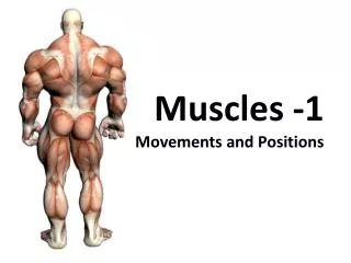
Muscles • Muscle is a general term for the tissues and organs of the body that produce force • The organ is sometimes referred to as whole muscle • There are three types of muscle tissue: • Skeletal (voluntary) • Cardiac (heart) • Smooth (hollow organs)
Whole Muscle • Whole muscle is an organ composed of skeletal muscle tissue wrapped in areolar connective tissue • Produces force when stimulated by a voluntary nerve fiber • Under conscious control • These are the muscles we think of as muscles
Whole Muscle Anatomy • A muscle has an origin and an insertion • Generally the more proximal attachment point is the origin and the distal attachment point is the insertion • Example: Biceps have origin at shoulder and insertion at elbow • Produce force by shortening
Whole Muscle Microanatomy • The muscle organ is composed of many cells called muscle fibers which are huge (each the size of a human hair) • Multinucleate due to being many cells fused together • These muscle fibers are arranged into bundles called fascicles
Muscle Fiber • A muscle fiber is a single cell composed of many fused cells • Most of the cell is filled with huge organelles called myofibrils • Myo- is the latin root for muscle • The myofibril is made up of many sarcomeres
Sarcomeres • The sarcomeres are the actual force-producing organelles of the cell • Composed essentially entirely of protein • This is why protein is essential to the diet of any athlete • Also why animal meat is full of protein
Muscle Stimulation • Muscles receive signals from motor neurons • These signals are transmitted by a neurotransmitter called acetylcholine • The signal is very brief so the muscle relaxes as soon as the signal is no longer being sent by the brain • These signals will be explored in more detail next week
T Tubule • The motor neuron’s signal reaches the outside of the muscle fiber and then is transmitted throughout the entire fiber through the T tubules • The signal is made up of sodium and potassium ions moving back and forth across the cell membrane • This is also known as an action potential • It moves much faster than diffusion
T Tubule and SR • When a neurotransmitter is released onto a muscle fiber, the electrochemical signal is transmitted through the T tubules through the entire fiber • As the signal moves through the T tubules, it stimulates the sarcoplasmic reticulum to release calcium • Modified version of the endoplasmic reticulum
Calcium? • Calcium ions are being constantly pumped into the SR • When the muscle is not being stimulated, all calcium ions are in the SR • When the muscle is stimulated, calcium is released but is still being pumped back into the SR • This means once the signal ends the muscle will stop contracting quickly • This requires a lot of constant ATP expenditure
Sarcomere • A sarcomere is made of two different kinds of protein fibers • Myosin filaments • Actin filaments • The myosin filaments have many myosin heads with two features: • They can attach to actin filaments • They try to bend
Sliding Filament Model • The myosin heads of the myosin filament attach to the actin filaments and attempt to move them together • There are two things that are required for this: • The actin can only be attached to when it has a calcium ion • The myosin head needs ATP to detach after it bends
Sliding Filaments • The actin filament has two molecules called troponin and tropomyosin • Together they block the myosin head from attaching to the actin filament • When a calcium ion attaches to the troponin, the tropomyosin is moved out of the way • The myosin head may now attach to the actin filament
ATP Usage • Clearly contracting our muscles requires energy • The actual use of the ATP is to detach the myosin head from the actin filament • Once the myosin head is detached it can reattach at a new point and pull again • Imagine if you had to stay in place and help play tug-of-war!
ATP Sources • All muscle cells have some free ATP when relaxed but this is depleted almost immediately during contraction • Once the ATP supply is depleted one of two things will happen: • More ATP must be produced rapidly • ATP will only be produced slowly by the mitochondria and the force produced will be less
Sources of ATP: • Creatine Phosphate (anaerobic) • Rapidly converted to an ATP, runs out after a few more seconds • Glycogen (anaerobic) • Stored glucose in the muscle fiber • Blood Glucose (aerobic) • Only absorbed and metabolized slowly • Fatty acids (aerobic) • Stored in muscle, metabolized slowly
ATP Depletion • Once the muscle’s main ATP supply is depleted, the available force produced is much lower • Fatigue • If all ATP is depleted, the muscle may lack the ATP supply to detach the myosin heads once the nerve signal ends • Cramp! • Once the muscle is relaxed, blood glucose and O2 are used to restore the ATP supply • Oxygen debt
Oxygen Debt • If more energy is used than can be produced aerobically, anaerobic production of energy can sustain activity • Produces by products such as lactic acid • Lactic acid can be reabsorbed and processed aerobically using more oxygen • This is why people pant and become winded if they have exerted themselves anaerobically
Rigor Mortis • When a person dies, they are initially limp • Once all ATP has been depleted from the cells, calcium begins to diffuse into the muscles • Calcium allows myosin head to attach to actin filament • Lack of ATP means the myosin head cannot be detached • Ultimately the entire body becomes incredibly stiff and will stay that way until the proteins physically break down from rotting (rigor mortis)