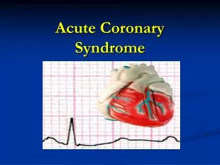
Acute coronary syndrome
790 likes | 2.47k Vues
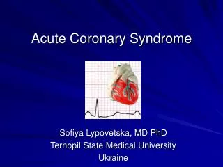
Acute coronary syndrome. Camille Ann L. Asuncion Case Presentation TMC IM-ER. General Data. TO 71 year old Male Filipino Roman Catholic Currently residing at Pasig City Brought to TMC ER last January 4, 2011 Informant and reliability: Self (good reliability). Chief Complaint.

Acute coronary syndrome
E N D
Presentation Transcript
Acute coronary syndrome Camille Ann L. Asuncion Case Presentation TMC IM-ER
General Data • TO • 71 year old • Male • Filipino • Roman Catholic • Currently residing at Pasig City • Brought to TMC ER last January 4, 2011 • Informant and reliability: Self (good reliability)
Chief Complaint • Chest Pain
History of Present Illness 8 hours PTC Chest pain sudden onset, substernal, nonradiating occurring at rest 20 minutes duration 10/10 in severity “more severe now than before” with associated diaphoresis No palpitations No difficulty of breathing No abdominal pain No fever Meds: IsorbideMononitrate (Imdur) 30 mg tab
History of Present Illness 3 hours PTC Recurrence of chest pain sudden onset, substernal, nonradiating occurring at rest 20 minutes duration 10/10 in severity with associated diaphoresis Headache diffuse, nonpulsating, nonradiating 5/10 in severity Meds: IsorbideMononitrate (Imdur) 30 mg tab
History of Present Illness Persistence of symptoms Rushed to TMC ER
Past medical history • (+) Diabetes Mellitus (~ 20 years) • Glimepiride (Norizec) 1 mg OD • Sitaglipin 50 g BID • (+) IHD s/p MI (2008) • Isosorbide-5-mononitrate (Imdur) 30 mg OD as needed • (+) PTB with pleural effusion (Nov. 2010) • s/p ultrasound guided thoracentesis (450 ml, right) • currently being treated with Rifampicin 150 mg, INH 75 mg, Pyrazinamide 400 mg, EthambutolHCl 275 mg (Quadtab) 3 tablets before breakfast, OD
Past medical history • No Hypertension • No asthma • No pneumonia • No allergies • No previous surgeries
Family history • (+) Diabetes Mellitus • mother • (+) asthma • paternal side • No Hypertension • No Pneumonia • No TB • No Heart disease
Personal and Social • Retired businessman • 20-pack year smoker • 1/2 pack per day • Occasional alcoholic beverage drinker • ~2-3 bottles of beer • Denies drug use/abuse
Review of Systems • Constitutional: no weight loss, no weakness, no fatigue • HEENT: no dizziness, no blurring of vision, no nosebleeds, no gum bleeding, no enlarged lymph nodes • Respiratory: cough, no dyspnea, no hemoptysis, no wheezing • Cardiovascular: no easy fatigability, no orthopnea, no syncope • Gastrointestinal: no nausea/vomiting, no change in bowel habits • Genitourinary: no dysuria, polyuria, no hematuria, no frequency • CNS: no seizure, no tremor • Muskuloskeletal: no muscle/joint pains, no joint swelling • Endocrine: no cold/heat intolerance
Physical examination • General Survey • Awake, cooperative, not in cardiorespiratory distress • Vital Signs • BP 120/70 HR 105 • RR 20 T 36.5 °C • Pulse Ox: 97% CBG 286 • Anthropometrics • Height 163 cm • Weight 65 kg • BMI = 24.4 kg/m2
Physical examination • Skin: No lesions. No rashes. No pigmentation or ulcers. • HEENT: Eyes:Anicteric sclera, pink palpebral conjunctiva. Ears: No tragal tenderness. Nose: No alar flaring. Septum midline. No discharge. No sinus tenderness. Mouth: Oral mucosa pink. Tongue midline. No tonsillopharyngeal congestion. • Neck: Supple. Trachea midline. Flat neck veins. No carotid bruits appreciated. Thyroid isthmus barely palpable, lobes not felt. • Lymph Nodes. No palpable cervical lymphadenopathies • Chest/Lungs: symmetric chest expansion, no visible retractions, decreased breath sounds on the right, no rales/crackles, no wheezes • Heart: adynamicprecordium, No lifets, heaves, thrills. Tachycardic, regular rhythm, Distinct S1, S2. No murmurs • Abdomen: Flat. No surgical scars, no visible veins or pulsations. Normoactive bowel sounds. No bruits. Tympanitic on percussion. Soft, no tenderness, no organomegaly. Liver edge not palpable. Spleen not palpable. • Extremities: No edema. No cyanosis. No clubbing. Full and equal pulses. No joint deformities. Good turgor (CRT <2 sec.)
Physical examination • Neurologic Examination • GCS 15 (E4 V5 M6) • Mental Status: Alert and cooperative, thought process coherent, oriented to person, place, and time. • Cranial Nerves: I – not tested; II, III, IV, VI – pupils are 2-3 mm, equally round and reactive to light and accommodations, full and equal extraocular movements, no nystagmus; V – temporal and masseter strength intact, bilateral facial sensation intact, corneal reflexes not tested; VII – bilateral facial movements intact, taste not tested; VIII – hearing equal for both right and left with finger wistling. X – gag reflex intact; XI – strength of sternocleidomastoid and trapezius muscles 5/5; XII – tongue midline. • Motor: Full range of motion in hands (5/5), wrists (5/5), elbows (5/5), shoulders (5/5), legs (5/5); no involuntary movements. • Cerebellar: Gait – Normal gait. Rapid alternating movements intact. Thumb-index finger pinch movements • Sensory: 100% intact sensation • No Kernig’s, No Babinski • Reflexes intact
Salient features • 71 year old • Male • Chest Pain • Sudden, substernal, heaviness, 20 min., at rest, with diaphoresis • 10/10 “more severe now than before” • (+) IHD s/p MI (2008) • (+) DM • 20-pack year smoker
Differential diagnoses • ACS • STEMI • NSTEMI • UNSTABLE ANGINA
Initial Diagnosis • Acute Coronary Syndrome • PTB 3 • DM II
Er diagnostics • ECG
Er diagnostics • ECG: possible inferior infarct • CBC • Cardiac Enzymes Hgb 110 Neut 0.68 Hct 0.33 Lymph 0.22 WBC 7.3 Mono 0.06 PC 304 Eosino 0.04 Hypochromic PT: Control vs. Patient 13.3 vs. 14.7 (12-14) % Activity 0.81 (0.7-1.31) INR 1.14 apTT: Control vs. Patient 32.2 vs. 30.8 (28-37) • Trop I (-) • CK-MB 14.54 (0-25) • CK-MM 11.83 (24-179) • CK-Total 26.37 (24-204)
Er diagnostics • Diagnostics • ECG • CBC • Cardiac Enzymes • Crea 0.79 mg/dl • Na 136 • K 4.4 • CXR: • Consider PTB with bronchiectatic changes, right upper lobe, unchanged. • Slightly progressing, pleural effusion, right
Er intervention • Supplemental Oxygen at 2 lpm via nasal cannula • Meds: • Aspirin (Aspec-EC) 80 mg tab OD • Clopidogrel (Plavix) 75 mg tab OD • Admitted to floors
Final Diagnosis • Unstable Angina • PTB 3 • DM II
Definition • Acute Coronary Syndrome • Any constellation of clinical symptoms that are compatible with acute myocardial ischemia • Spectrum of disease, due to an imbalance of myocardial oxygen demand and supply
Definition complete obstruction of a coronary artery damage/necrosis of the full thickness of the heart muscle Partial obstruction of a coronary artery damage/necrosis of the partialthickness of the heart muscle Vs. NSTEMI : severity of ischemia to cause sufficient myocardial damage; Cardiac marker (-)
diagnosis • Risk Factors • Modifiable • Smoking • Hypercholesterol • Hypertension • Obesity • Diabetes Mellitus • Physical Inactivity • Non-modifiable • Age • Male • Family History of early MI • <50 y/o males • <55 y/o females • Known CAD
diagnosis • Signs and Symptoms • Prolonged (usually > 30 minutes) constricting, crushing, squeezing pain • retrosternal, radiating to left chest, left arm • can be epigastric • sense of indigestion • Nausea/vomiting (inferior > anterior MI) • Palpitations • Diaphoresis • Sense of “impending doom” • *may be asymptomatic in diabetics
diagnosis • “high likelihood patient” • Established CAD by angiography • History of CABG or PCI • History of MI, CHF • Multiple CAD risk factors
pathophysiology 1. Endothelial dysfunction • Hypercholesterolemia LDL particles oxidative modification inflammatory response leukocyte adhesion molecules monocyte adhesion and migration 2. Fatty Streak • Phagocytes ingest lipids foam cells 3. Advanced, Complicated Lesion • Migration of smooth muscle cells - accumulation fibrous cap
pathophysiology 4. Unstable Fibrous Plaque • Lesion expansion apoptosis, necrosis 3. Plaque Rupture with thrombus • Clot overwhelms fibrinolytic mechanisms Rupture of fibrous cap thrombosis
Diagnostics • Exercise Stress Testing • Walk on treadmill at increasing levels of difficulty • Target heart rate = 85% maximum of age • (+) CAD • ST elevation • ST depression > 1mm in multiple leads • Decreased BP • Failure to exercise more than 2 minutes due to symptoms
Diagnostics • ECG • ST elevation • Inferior (II, III, aVF) • Anteroseptal (V1, V2, V3) • Lateral (V4, V5, V6) • ST depression • Posterior (V1, V2) • T wave inversion • Ischemia • Manifestations can vary depending on its location in the heart • Anterior – LAD • Posterolateral – Circumflex
Diagnostics • Cardiac Markers • Troponin I (within 3 hours ~1 week) • Sensitive and specific • CK-MB • CK-MM • CK-Total
treatment • Initial Treatment for all ACS (UA/NSTEMI) • Anti-ischemic • Oxygen • NTG • Morphine • Beta blockers • Decrease cardiac oxygen demand • Antiplatelet and anticoagulation • Aspirin • Clopidogrel • Heparin • Unfractionated • Low molecular weight • GP IIb/IIIa Inhibitors
treatment • *Thrombolytics are not used in UA or NSTEMI because in 60-80% the infarcted artery is not occluded. • STEMI • Early revascularization with thrombolytics • Streptokinase, Urokinase, etc. • and/or cardiac catheterization and stent • elevated troponin • Recurrent chest pain despite medical therapy • CHF • Positive stress test • Left ventricular EF< 40% • Sustained ventricular tachycardia • Cardiac stent within 6 months
prognosis • TIMI Risk Score • Age >= 65 years • >= 3 risk factors for CAD • Prior coronary stenosis >= 50% • Presence of ST segment deviation on admission ECG • At least 2 anginal episodes in last 24 hours • Elevated serum cardiac biomarkers • Use of aspirin prior seven days
Public health and Prevention • Promote Healthy Lifestyle • DIET modification • Smoking cessation • Diabetes management • Hypertension control
Acute coronary syndrome Camille Ann L. Asuncion Case Presentation TMC IM-ER