Download
1 / 53
600 likes | 1.13k Vues
The sensory system and pain syndromes. Vth year, dentistry, 30 . 09 .200 8 Department of Neurology Semmelweis University. Sensory system. Receptors: - specialised (smell, vision, hearing, taste - visceral (viscera, smooth muscle - unconscious or autonomic)

E N D
The sensory system and pain syndromes Vth year, dentistry,30.09.2008 Department of Neurology Semmelweis University
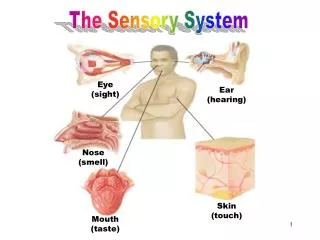
Sensory system Receptors: - specialised (smell, vision, hearing, taste - visceral (viscera, smooth muscle - unconscious or autonomic) - somatic (skin, striated muscle, joints)
Cutaneous receptors Muscle, tendon receptors
The sensory system Spinothalamic system (tractus spinothalamicus) exteroceptive sensation) :pain temperature light touch Dorsal column pathway ( lemniscus medialis) “conscious” proprioception: joint position vibration deep pressure two point discrimination graphaesthesia !stereoaesthesia ! Dorsal and ventral spinocerebellar pathway “unconscious” proprioception
Pain Nociceptors: -Unimodal: mechanoreceptors, thinly myelinated fiber -Bimodal: cold + mechanoreceptors, thinly myelinated and unmyelinated fibers warm+mechanoreceptors -Polymodal:warm-mechano-chemical receptors, unmyelinated fibers
Spinothalamic system Pain perception C fibers: thin, unmyelinated A delta: thinly myelinated Temperature A delta: thinly myelinated
Neuropathic pain primary lesion or dysfunction of CNS (peripheral or central)1 Nociceptiv pain caused by tissue damage (bones,joints, tendons, muscles, skin, viscera)2 Mixed origin of pain - manifestations • Peripheral: • Postherpetic neuralgia • Trigeminal neuralgia • Polyneuropathy in diabetes mell. • Posttraumatic neuropathy • Central: • Poststroke pain • Description:2 • Burning • Tickling, itching,pins and needles • Hypersensitivity to touch/cold • Low back pain with radiculopathy • Cervical pain with radiculopathy • Cancer pain • Carpal tunnel syndrome • inflammation • fractures • osteoarthritis. • Postoperativ visceral pain • Description:2 • smarting • Sharp • Pulsating, throbbing 1. International Association for the Study of Pain. IASP Pain Terminology. 2. Raja et al. in Wall PD, Melzack R (Eds). Textbook of pain. 4th Ed. 1999.;11-57
Dorsal column pathway/lemniscus medialis Proprioceptiv modalities: pressure, vibration, joint positiontwo points discrimination, graphaesthesia ! stereoaesthesia ! Type of fibers: thick, myelinated fibers (Aα, I, II)
Sensory disturbances Positive symptoms: • Pain • Hyperaesthesia:increased sensitivity to any stimulus • Hyperalgesia: increased sensitivity to a painful stimulus • Hyperpathia: increased sensitivity with increasing pain threshold to repetitive stimulation • Paraesthesia:“pins and needlessensation”, “burning feeling” • Dysaesthesia: inappropriate sensation to a stimulus • Allodynia: pain provoked by a non-painful stimulus
Sensory disturbances Negative symptoms: • Hypoalgesia: reduced sensitivity to a painful stimulus • Hypoesthesia: reduced sensitivity to any stimulus • Analgesia: absent sensitivity to a painful stimulus • Anaesthesia: absent sensitivity to any stimulus
Examination of the sensory system 1. Special standpoints: • “Subjective “ examination • Requires good cooperation on the patient`s side. • Allows accurate localisation of the pathology. • Preliminary diagnosis is needed. Examine according to the expected damage ! • Most often we compare different parts of the body. • Do not tell the patient what should be felt ! • The patient should not see the examined part of the body ! • “Subjective” sensory disturbance ( pain, paraesthesia ) is not necessarily accompanied by “objective” sensory disturbance (hypaesthesia, anaesthesia )
Examination of the sensory system 2. Pain: pin prick, tooth picks Light touch: use a wisp of cotton wool ! Temperature:use cold (5-10 0C)/or hot (40-45 0C) test tubes ! -Instruct the patient to reply:“Tell me if you feel the stimulus ! Name the area stimulated !” “Is it equal on both sides? -Map out the extent of abnormality by moving from the abnormal to the normal area (“Tell me if sensation changes!”) Joint position / motion: -Hold the sides of the patient’s finger !Move it up and down at random !Ask to specify the direction of movement ! Vibration: -Place a vibrating tuning fork on a bony prominence ( ankle, knee, processus spinosus, processus styloideus radii et ulnae, elbow, clavicula)
Examination of the sensory system 3. • Two point discrimination: -The ability to discriminate two blunt points when applied simultaneously. (3-5 mm on the finger, 4-7 cm on the trunk) • Sensory inattention (perceptual rivalry) -The ability to detect sensory stimuli applied simultaneously on both limbs. -Subdominant parietal lobe, associative areas • Stereoaesthesia - An object is placed in the patient’s hand. - Ask patient to describe its size, shape, surface, material ! - Stereoanaesthesia:disturbance of the sensory afferent tracts.
Examination of the sensory system 4. • Astereognosis. -Inability to identify an object by palpation -The primary sense data being intact -Lesion of the opposite hemisphere, postcentral gyrus • Tactile agnosia: -The patient is unable to recognize an object by touch in both hands -Disorder of perception of symbols. -Lesion of the dominant parietal lobe, associative areas • Graphaesthesia - The ability to recognize numbers or letters traced out on the palm.
Examination of the sensory system • Nerve conduction studies: sensory antidrom neurography median nerve, ulnar nerve • Somatosensory evoked potentials (SEP) median nerve, tibial nerve
Peripheral nerve, Polyneuropathies Peripheral nerve: according to the distribution area of the affected nerve Polyneuropathies: symmetrical sensory disturbance in stocking/glove like distribution, more pronounced distally Sensory disturbance usually starts on the toes, gradually spreads higher, rarely above the knee; later on the hands
Spinal ganglion segmental, localised to dermatomes
Root damage • Sensory disturbance and pain according to the dermatome (variability!) • Anaesthesia does not develop because of overlapping dermatomes C7 S1
Syringomyelia • spinothalamic fibers crossing at cervical level are affected first • dissociated sensory loss: temperature, pain disturbance on both hands
Cranial structures - pain • skull • cervical spine • eyes • ears • nose, sinuses • teeth • temporomandibular joint
HeadachePathways PAG:periaqueductal gray matter LC: locus ceruleus TG:trigeminal ganglion DRG:dorsal root ganglion
Taking a headache history • Age of onset ? • Duration of complaint ? • Time pattern Continuous or transient ? Frequency and duration of each headache ? • Site ? • Intensity, quality ? • Associated phenomena ? • Aggravating and relieving factors ?
Headache - „danger signals” „Danger signals”: • sudden onset of new, severe headache • onset of headache after exertion, straining, coughing or sexual activity • progressively worsening headache • any abnormality on neurological examination • systemic features: fever, arthralgia • onset of first headache after the age of 50 years Refer to specialist: • Cooperating patient – ineffective treatment • Chronic daily headache – drug abuse, dependency • Severe anxiety , depression • Severe comorbid diseases
International classification of headache disorders Primary headaches: 1. Migraine 2. Tension-type headache 3. Cluster headache and other trigeminal autonomic headaches 4. Other primary headaches Secondary headaches: 5. Posttraumatic (head/neck trauma) 6. Vascular disorder (cranial/cervical) 7. Non vascular intracranial disorder 8. Substance abuse/ withdrawal 9. Infection 10 Disorder of homeostasis 11.Disorder of facial/cranial structures 12.psychiatric disorder Cranial neuralgias, central and primary facial pain : 13.Cranial neuralgias and central causes of facial pain 14.Other headache International Headache Society. ICHD-II. Cephalalgia 2004; vol 24: suppl 1.
Migraine without aura A) n 5 B) 4 - 72 h C) 1. 2. 3. ++ / +++ 4. D) 1. + 2. E) normal 2/4 / 1/2
Phases of migraine Blau, Lancet, 1992
Woods et al. 1994 Migraine and „spreading depression”
Trigemino - vascular system Lancet Neurology 2002;1:251-257.
migraine - treatment • Acute: • - Non specific:analgesics, NSAID, antiemetics • - Specific:ergotamine, dihydroergotamine, triptans • Preventive (prophylactic): • - Episodic: if there is a trigger for a limited time ( menses) • - Chronic:decrease the frequency independently of triggers • Migrén gyógyszeres kezelésének protokollja, • Magyar Fejfájás Társaság,2003
Tension type headache: criteria A. n > 10 B. 30 min < duration of pain< 7 nap C. 2/4 + / ++ D. 2/2 E. normal /
Convergence and sensitisation in the trigeminal nuclei Thalamus DRN, LC pia / dura vessels art. temp. Trigeminus nucleus caudalis masticatory muscle Brainstem and spinal cord
Tension type headache - treatment Acute treatment: analgesics NSAID + antiemetics, coffein Preventive treatment:tricyclic AD SSRI valproat (?) Complex treatment: pharmacological treatment psychotherapy relaxation physiotherapiy (Not massage!)
Cluster headache: criteria A) n 5 B) +++ 30-180 min D) Frequency = 1/2 ->50 E) normal C) 1/4
Cluster headache: treatment Acute treatment: oxygen (7 l/min, 10 perc) sumatriptan sc. inj. ergotamine indomethacin supp. Preventive treatment: verapamil (360 mg/day) valproate (600-1500 mg /day) infiltration of occipital nerve ? methysergide, pizotifen ? lithium (chronic cluster!) corticosteroids/dihydroergotamine Surgical ?
International classification of headache disorders Secondary headaches: 5. Posttraumatic (head/neck trauma) 6. Vascular disorder (cranial/cervical) 7. Non vascular intracranial disorder 8. Substance abuse/ withdrawal 9. Infection 10 Disorder of homeostasis 11.Disorder of facial/cranial structures 12.psychiatric disorder Primary headaches: 1. Migraine 2. Tension-type headache 3. Cluster headache and other trigeminal autonomic headaches 4. Other primary headaches Cranial neuralgias, central and primary facial pain : 13.Cranial neuralgias and central causes of facial pain 14.Other headache International Headache Society. ICHD-II. Cephalalgia 2004; vol 24: suppl 1.
Trigeminal neuralgia V/1 V/2 V/1 V/1 V/3 V/3 C2/3
Trigeminal neuralgia • Prevalence: 10-20 / 100 000 population • female/male : 1.6 • age of onset: > 50 years (90%) • site: most frequently V/2,3 < 5 % V/1 division ~ 10 % all the three division ~ 5 % bilateral Features: • placebo effect 0 -1 % ! • trigger zone 90 % • refracter phase • spontanous remission ~ 50 %, < 6 months • „pretrigeminal neuralgia”
Trigeminal neuralgia • Symptomatic • pain as described before • persistence of aching between paroxysm • sensory impairment or other neurological deficit • causative lesion , other than vascular compression Classical/Idiopathic • duration < 2 minutes • affecting one/more divisions • sudden onset • severe, sharp,stabbing pain • precipitated from trigger areas • patiens is pain free between paroxysm • no neurological deficit • no causative lesion
Trigeminal neuralgia mouth -ear zone, 60% nose - orbit zone, 30 %
Peripheral aetiology -central pathogenesis chronic irritation of trigeminal nerve division focal demyelinisation ectopic action potentials decrease of segmental inhibition paroxysmal discharge of LTM interneurons of nucleus oralis n.V paroxysmal discharge of WDR neurons of nucleus caudalis n.V attack of trigeminal neuralgia
Trigeminal neuralgia • differential diagnosis • examinations: anamnesis physical examination Rtg otology dental surgery ophthalmology brain MR trigeminal SEP psychology „diagnostic blockade”
Trigeminal neuralgia • If it is possible determine causative lesion, treat it • Pharmacological treatment - antiepileptics - muscle relaxants - tranquillants • TENS? • surgery
Trigeminal neuralgia-pharmacological treatment • Carbamazepine 400-1200 mg/day • Phenytoin 300-600 mg/day • Valproate 500-2000 mg/day • L baclofen 40-80 mg/day • Clonazepam 2-8 mg/day • Pimozide 4-12 mg/day • Tiapridal 300-600 mg/day • Gabapentin up to 3600 mg/day • Pregabalin ?
Trigeminal neuralgia - phamacological treatment • start with small dose, increase gradually • prefer combination • blood counts, hepatic, renal function tests are needed • monitoring of complaints is important • timing of discontinuation (after pain free for 8 weeks) • gradual tapering is necessary • 30 % of patients fail to respont to medical therapy
Trigeminal neuralgia – surgical management • Peripheral nerve blockade • Percutanous radiofrequency trigeminal thermocoagulation (Sweet, Wespic, 1974) • Retrogasserian Glycerol injection(Hakansson, 1981) • Radiosurgery - gamma knife • Microvascular decompression (Gardner 1966, Janetta 1967, MéreiFT 1973) Janetta: 85 % vascular compression a. cerebelli superior V/2,3 a. cerebelli inferior anterior V/1 pain free : 80%, mortality: 0.5 %
neuralgias Glossopharyngeal neuralgia: classical/symptomatic - pain in the tonque, tonsillar fossa, angle of the jaw, ear - peritonsillar abscess, oropharyngeal carcinoma ! Nervus intermedius neuralgia - posterior wall of the auditory canal - herpes zoster oticus ! Superior laryngeal neuralgia Nasociliary neuralgia Supraorbital neuralgia Occipital neuralgia - greater or lesser occipital nerves - cervical spine !