Download
1 / 1
10 likes | 145 Vues
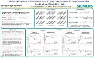
Weights and Topology: A Study of the Effects of Graph Construction on 3D Image Segmentation. Leo Grady and Marie-Pierre Jolly. Leo.Grady@Siemens.com, Marie-Pierre.Jolly@Siemens.com . Gaussian weights:. Reciprocal weights:. Histogram weights:. 6-connected. 10-connected. 26-connected.

E N D
Weights and Topology: A Study of the Effects of Graph Construction on 3D Image Segmentation Leo Grady and Marie-Pierre Jolly Leo.Grady@Siemens.com, Marie-Pierre.Jolly@Siemens.com Gaussian weights: Reciprocal weights: Histogram weights: 6-connected 10-connected 26-connected Weighting Random Walker Graph Cuts Graph Cuts Random Walker Department of Imaging and Visualization Siemens Corporate Research, Princeton Weighting types Topology types Main Idea Many graph-based segmentation algorithms are popular: Random walker, graph cuts, normalized cuts, fuzzy connectivity, etc. All of these algorithms use the same 3D graph topologies and intensity weighting functions. This work studies the effects of several common topologies/weightings on the segmentation performance of two graph-based segmentation algorithms on 62 3D CT datasets compared to ground truth segmentations. w is edge weight, g is image intensity, p is probability of intensity, β is a free parameter Results Data used Topology 62 3D medical datasets containing a single segmentation target were manually segmented by a clinical practitioner. Each volume was also given manually-placed foreground and background seeds by the same clinical practitioner that provided the manual segmentation. The data contained a range of segmentation targets including tumors, lymph nodes, cysts and other lesions. The data was acquired using different Siemens CT scanners with different reconstruction kernels. The clinical input (ground truth and seeds) was given by different clinical partners. The datasets we used for segmentation were typically cropped from larger data acquisitions and ranged in size from roughly (40 × 40 × 40) to (128 × 128 × 128). Most datasets had different numbers of voxels in each dimension and had a greater spacing between axial slices than within the slice. All of the data acquisitions were axial scans. The XY-plane was chosen to correspond to an axial slice.