Download
1 / 19
190 likes | 394 Vues
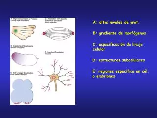
A: altos niveles de prot. B: gradiente de morfógenos. C: especificación de linaje celular. D: estructuras subcelulares. E: regiones específica en cél. o embriones. Proteínas que reconocen al mRNA target. PCR degradativa.

E N D
A: altos niveles de prot. B: gradiente de morfógenos C: especificación de linaje celular D: estructuras subcelulares E: regiones específica en cél. o embriones
The localization of RNAs at the vegetal cortex in Xenopus oocytes is a complex process, involving at least two different pathways. The early, or messenger transport organizer (METRO), pathway, localizes RNAs such as Xlsirts, Xcat2 and Xwnt11 during stages 1 and 2 of oogenesis, while the late pathway localizes RNAs such as Vg1 during stages 2–4. We demonstrate that the onset of Vg1 localization is characterized by its microtubule-independent binding to a subdomain of the endoplasmic reticulum (ER). The formation of this unique ER structure is intimately associated with the movement of the mitochondrial cloud toward the vegetal cortex. In addition, we demonstrate that the mitochondrial cloud contains a -tubulin-positive structure that may function as a microtubule organizing center for establishing microtubule tracks for Vg1 localization. These data, support, although they do not prove, a model in which the development of the late pathway machinery relies upon the prior functioning of the early pathway.
Fig. 7. Model for the establishment of the late pathway. The Vg1 mRNA (red dots) is distributed throughout the cytoplasm while Xlsirts, Xcat2 and Xwnt11 are within the METRO (blue) in stage 1 oocytes (A). Later during stage 1 we detect a cap of ER material associated with the side of the migrating cloud closest to the GV (orange) (B). By stage 2 a portion of the ER forms a wedge-shaped structure that occupies the channel created by the migrating mitochondrial cloud (C). During this time we detect Vg1 mRNA co-localizing with this subdomain of the ER. This association is not microtubule dependent. Xlsirts, Xcat2 and Xwnt11 are already localized as a disk at the apex of the vegetal pole (blue). By stage 3 Vg1 mRNA is translocating to the vegetal cortex, utilizing microtubules. There are two possible means by which this occurs. The first (D) is that Vg1 remains directly associated with the ER (possibly through the vera protein; Deshler et al., 1997) and ER vesicles are moved along microtubules (green). Alternatively, (D') the ER structure serves as a matrix for the laying down of the microtubules and the Vg1 mRNA translocates along the microtubules, perhaps binding to the microtubules through the 69-kDa Vg1 binding protein (Schwartz et al., 1992). The green spot within the mitochondrial cloud represents the putative remnants of the centrosome that may function as an MTOC at the vegetal cortex to produce the microtubules used to translocate Vg1. Note that the model only depicts a portion of the microtubules present in stage 3 oocytes.